Showing posts with label bone marrow. Show all posts

Tuesday, April 3, 2012

Three Types of Transplants For Cancer Treatment

Transplants have played a vital role in cancer treatment in the past few years. Many leukemia and lymphoma patients have benefited from this procedure.  Many lab tests have to be performed before becoming a candidate to receive a transplant. There are three types of transplants that are available in the medical community.

Autologous

Autologous HSCT requires the extraction (apheresis) of haematopoietic stem cells (HSC) from the patient and storage of the harvested cells in a freezer. The patient is then treated with high-dose chemotherapy with or without radiotherapy with the intention of eradicating the patient's malignant cell population at the cost of partial or complete bone marrow ablation (destruction of patient's bone marrow function to grow new blood cells). The patient's own stored stem cells are then returned to his/her body, where they replace destroyed tissue and resume the patient's normal blood cell production. Autologous transplants have the advantage of lower risk of infection during the immune-compromised portion of the treatment since the recovery of immune function is rapid. Also, the incidence of patients experiencing rejection (graft-versus-host disease) is very rare due to the donor and recipient being the same individual. These advantages have established autologous HSCT as one of the standard second-line treatments for such diseases as lymphoma. However, for others such as Acute Myeloid Leukemia, the reduced mortality of the autogenous relative to allogeneic HSCT may be outweighed by an increased likelihood of cancer relapse and related mortality, and therefore the allogeneic treatment may be preferred for those conditions. Researchers have conducted small studies using non-myeloablative hematopoietic stem cell transplantation as a possible treatment for type I (insulin dependent) diabetes in children and adults. Results have been promising; however, as of 2009 it was premature to speculate whether these experiments will lead to effective treatments for diabetes.

Allogeneic

Allogeneic HSCT involves two people: the (healthy) donor and the (patient) recipient. Allogeneic HSC donors must have a tissue (HLA) type that matches the recipient. Matching is performed on the basis of variability at three or more loci of the HLA gene, and a perfect match at these loci is preferred. Even if there is a good match at these critical alleles, the recipient will require immunosuppressive medications to mitigate graft-versus-host disease. Allogeneic transplant donors may be related (usually a closely HLA matched sibling), syngeneic (a monozygotic or 'identical' twin of the patient - necessarily extremely rare since few patients have an identical twin, but offering a source of perfectly HLA matched stem cells) or unrelated (donor who is not related and found to have very close degree of HLA matching). Unrelated donors may be found through a registry of bone marrow donors such as the National Marrow Donor Program. People who would like to be tested for a specific family member or friend without joining any of the bone marrow registry data banks may contact a private HLA testing laboratory and be tested with a mouth swab to see if they are a potential match. A "savior sibling" may be intentionally selected by preimplantation genetic diagnosis in order to match a child both regarding HLA type and being free of any obvious inheritable disorder. Allogeneic transplants are also performed using umbilical cord blood as the source of stem cells. In general, by transplanting healthy stem cells to the recipient's immune system, allogeneic HSCTs appear to improve chances for cure or long-term remission once the immediate transplant-related complications are resolved.
A compatible donor is found by doing additional HLA-testing from the blood of potential donors. The HLA genes fall in two categories (Type I and Type II). In general, mismatches of the Type-I genes (i.e. HLA-A, HLA-B, or HLA-C) increase the risk of graft rejection. A mismatch of an HLA Type II gene (i.e. HLA-DR, or HLA-DQB1) increases the risk of graft-versus-host disease. In addition a genetic mismatch as small as a single DNA base pair is significant so perfect matches require knowledge of the exact DNA sequence of these genes for both donor and recipient. Leading transplant centers currently perform testing for all five of these HLA genes before declaring that a donor and recipient are HLA-identical.
Race and ethnicity are known to play a major role in donor recruitment drives, as members of the same ethnic group are more likely to have matching genes, including the genes for HLA.

Synogeneic 

Syngeneic bone marrow transplantation is a procedure in which a person receives bone marrow donated by his or her healthy identical twin.  This is more of a specific term for a for the procedure above.


For more information:
http://www.cancer.gov/cancertopics/factsheet/Therapy/bone-marrow-transplant

http://en.wikipedia.org/wiki/Syngeneic_bone_marrow_transplantation


Thursday, March 22, 2012

Myeloproliferative Dimyeloproliferative disorders

Myeloproliferative disease or MPD deals with  a group of diseases of the bone marrow in which excess cells are produced.


Myeloproliferative disorders is the name for a group of conditions that cause blood cells -- platelets, white blood cells, and red blood cells -- to grow abnormally in the bone marrow. Though myeloproliferative disorders are serious, and may pose certain health risks, people with these conditions often live for many years after diagnosis. The prognosis largely depends on the type of disorder.
Myeloproliferative disorders include:
  • Polycythemia vera -- occurs when the bone marrow produces too many blood cells, especially red blood cells. More than 95% of people with polycythemia vera carry the blood mutation JAK2V617F.
  • Essential thrombocytosis -- occurs when the body produces too many platelet cells, which help blood to clot. Clots can block blood vessels leading to heart attack or stroke.
  • Primary or idiopathic myelofibrosis, also known as myelosclerosis -- occurs when the bone marrow produces too much collagen or fibrous tissue in the bone marrow. This reduces bone marrow's ability to produce blood cells.
  • Chronic myelogenous leukemia (CML) -- cancer of the bone marrow that produces abnormal granulocytes, a type of white blood cell, in the bone marrow.

Signs and Symptoms:

Many people with myeloproliferative disorders have no symptoms when their doctors first make the diagnosis. One symptom shared by all myeloproliferative disorders, with the exception of essential thrombocytosis, is an enlarged spleen. An enlarged spleen can cause abdominal pain and a feeling of fullness.

Many times, especially in the early stages, myeloproliferative disease does not have symptoms. When it does have signs, they vary from person to person. If you have symptoms, they may include:
  • Headache
  • Fatigue
  • Shortness of breath
  • Easy bruising or bleeding
  • Petechiae (tiny red spots under the skin)
  • Unexplained weight loss
  • Night sweats
  • Fever
Specific disorders in which the bone marrow produces too many cells have similar and yet different symptoms.

Some signs and symptoms of the different types of myeloproliferative disorders include:
Polycythemia vera
  • Fatigue, general malaise
  • Trouble breathing
  • Intense itching after bathing in warm water
  • Stomachaches
  • Purple spots or patches on the skin
  • Nosebleeds, gum or stomach bleeding, or blood in the urine
  • Throbbing and burning pain in the skin, often with darkened, blotchy areas
  • Headache and problems with vision
  • High blood pressure
  • Blockage of blood vessels. This may cause heart disease, stroke, or gangrene (tissue death) of the arms and legs.
Essential thrombocytosis
  • Heart attack or stoke
  • Headache
  • Burning or throbbing pain, redness, and swelling of the hands and feet
  • Bruising
  • Gastrointestinal bleeding or blood in the urine
Primary myelofibrosis
  • Fatigue, general malaise
  • Trouble breathing
  • Anemia
  • Weight loss
  • Fever and night sweats
  • Abnormal bleeding
Chronic myelogenous leukemia (CML)
  • Fatigue, general malaise
  • Weight loss or loss of appetite
  • Fever and night sweats
  • Bone or joint pain
  • Heart attack or stroke
  • Trouble breathing
  • Gastrointestinal bleeding
  • Infection

Laboratory Tests

Complete blood count (CBC) and differential CBCs and differentials are the most frequently ordered tests used to help diagnose and monitor MPDs. Often ordered as part of a yearly physical exam, they are routine tests that count the number and relative proportion of each of the different types of cells in your blood stream. They give your doctor information about the size, shape, and relative maturity of the blood cells present in your blood at that moment.
CBCs and differentials can be used to detect WBC, RBC, and platelet increases, decreases, and abnormalities. They can help determine their severity, diagnose their cause, monitor the course of a disease, and monitor the response to treatment.
With polycythemia vera, increased RBCs, platelets, and sometimes WBCs, may be seen. With myelofibrosis, immature granulocytes and misshapen immature teardrop-shaped red blood cells are often seen, and WBC and RBC numbers are often decreased. With thrombocythemia, greatly increased numbers of platelets are seen along with abnormally large platelets, platelet clumps, and fragments of megakaryocytes.
Irregularities in cell counts may be due to MPDs, but they may also be due to a variety of other temporary or chronic conditions. Other testing is usually done to confirm or rule out the diagnosis of an MPD.
Bone marrow aspiration/biopsyIf a doctor suspects a bone marrow disorder, he may order a bone marrow aspiration or biopsy to collect a small sample of marrow. When a specialist (a pathologist, oncologist, or hematologist) examines the bone and fluid from the bone marrow sample under the microscope, he can see the number, size, and shape of precursor cells (blasts), red and white blood cells, and megakaryocytes (platelet precursors). He can determine the proportions of mature and immature cells, see any overgrowth of fibrous tissue, and detect any cancer cells from cancers that may have spread to the marrow. Most bone marrow disorders can be diagnosed during this examination.

ABGs (Arterial blood gases) - This test measures the amount of gases in your arterial blood and may be done when polycythemia vera is suspected. Low levels of oxygen are associated with secondary polycythemia.
Erythropoietin is a hormone that stimulates the bone marrow to produce RBCs. With primary polycythemia, erythropoietin levels will be very low or absent, but with secondary polycythemia they will be normal or high.
Genetic testing is sometimes used in suspected chronic myelogenous leukemia to check for the presence or absence of a Philadelphia (Ph') chromosome or a bcr-abl translocation (see BCR ABL) and in suspected polycythemia vera, myelofibrosis, and essential thrombocythemia for the presence or absence of JAK2 mutations, a gene associated with marrow cell production.

                                        Video of Bone Marrow Biopsy


              This video is a lecture about a very common MPD  which is polycythemia vera.  It is seen frequently in persons who live in high altitudes.

For more information then check the following websites:

http://www.umm.edu/altmed/articles/myeloproliferative-disorders-000114.htm or

http://labtestsonline.org/understanding/conditions/myelopro-disorders?start=2
                                      

Friday, March 9, 2012

Acute Monocytic Leukemia

There are several types of white blood cells in our blood.  Above is a chart of the different kinds which include lymphocytes, monocytes, basophils, neutrophils and eosinophils. Any time any of these cell are produced rapidly from the bone marrow and seen in immature stages in the peripheral blood system then this is usually caused by cancer and is determined to be a leukemia.

In this article we will be addressing acute monocytic leukemia.  Acute stage of any leukemia is when the bone marrow produces an abnormal amount of premature white cells that can be seen in our blood system through a microscope.


Notice how large these monocytes are seen in the peripheral blood under a microscope after a smear has been made on a glass slide and then stained with Wrights. These are called white cells in the blast stage which is very immature.  Notice the center nucleus is very large. A normal monocyte nucleus is small and condensed.  The outer cytoplasm has vacuoles (holes).

Acute myeloid leukemia (AML) is one of the most common types of leukemia among adults. This type of cancer is rare under age 40. It generally occurs around age 60. (This article focuses on AML in adults.)
AML is more common in men than women.
Persons with this type of cancer have abnormal cells inside their bone marrow. The cells grow very fast, and replace healthy blood cells. The bone marrow, which helps the body fight infections, eventually stops working correctly. Persons with AML become more prone to infections and have an increased risk for bleeding as the numbers of healthy blood cells decrease.
Most of the time, a doctor cannot tell you what caused AML. However, the following things are thought to lead to some types of leukemia, including AML:
  • Certain chemicals (for example, benzene)
  • Certain chemotherapy drugs, including etoposide and drugs known as alkylating agents
  • Radiation
Problems with your genes may also play a role in the development of AML.
You have an increased risk for AML if you have or had any of the following:

Exams and Tests

The doctor will perform a physical exam. There may be signs of a swollen spleen, liver, or lymph nodes.
A complete blood count (CBC) shows anemia and a low number of platelets. A white blood cell count (WBC) can be high, low, or normal.
Bone marrow aspiration will show if there are any leukemia cells.
If your doctor learns you do have this type of leukemia, further tests will be done to determine the specific type of AML. There are eight subtypes of AML. They range from M0 to M7, based on which blood cells are abnormal.

Treatment

Treatment involves using medicines to kill the cancer cells. This is called chemotherapy. But chemotherapy kills normal cells, too. This may cause side effects such as excessive bleeding and an increased risk for infection. Your doctor may want to keep you away from other people to prevent infection.
Other treatments for AML may include:
  • Antibiotics to treat infection
  • Bone marrow transplant or stem cell transplant after radiation and chemotherapy
  • Red blood cell transfusions to fight anemia
  • Transfusions of platelets to control bleeding
Most types of AML are treated the same way. However, a form of AML called acute promyelocytic leukemia (APL) is treated with a medicine called all-trans retinoic acid (ATRA). This medicine helps leukemia cells grow into normal white blood cells.
The drug arsenic trioxide is for use in patients with APL who do not get better with ATRA or chemotherapy.

Support Groups

See:

Outlook (Prognosis)

When the signs and symptoms of AML go away, you are said to be in remission. Complete remission occurs in most patients.
With treatment, younger patients with AML tend to do better than those who develop the disease at an older age. The 5-year survival rate is much lower in older adults than younger persons. Experts say this is partly due to the fact that the body of a younger person can better tolerate strong chemotherapy medicines.
If the cancer does not come back (relapse) within 5 years of the diagnosis, you are considered permanently cured.

Possible Complications

Complications of AML and cancer treatment include severe infections and life-threatening bleeding. Sometimes, the cancer comes back (relapses) after treatment.

                      This video will explain about leukemias in acute and chronic stages.

For more information:  http://www.nlm.nih.gov/medlineplus/ency/article/000542.htm

Monday, February 20, 2012

When Does A Patient Need A Stem Cell Transplant?

Let's first give a definition of a stem cell.
Stem cells have the remarkable potential to develop into many different cell types in the body during early life and growth. In addition, in many tissues they serve as a sort of internal repair system, dividing essentially without limit to replenish other cells as long as the person or animal is still alive. When a stem cell divides, each new cell has the potential either to remain a stem cell or become another type of cell with a more specialized function, such as a muscle cell, a red blood cell, or a brain cell.
Stem cells are distinguished from other cell types by two important characteristics. First, they are unspecialized cells capable of renewing themselves through cell division, sometimes after long periods of inactivity. Second, under certain physiologic or experimental conditions, they can be induced to become tissue- or organ-specific cells with special functions. In some organs, such as the gut and bone marrow, stem cells regularly divide to repair and replace worn out or damaged tissues. In other organs, however, such as the pancreas and the heart, stem cells only divide under special conditions.

 
A stem cell transplant is the infusion of healthy stem cells into your body. A stem cell transplant may be necessary if your bone marrow stops working and doesn't produce enough healthy stem cells. A stem cell transplant can help your body make enough healthy white blood cells, red blood cells or platelets, and reduce your risk of life-threatening infections, anemia and bleeding.
Although the procedure to replenish your body's supply of healthy blood-forming cells is generally called a stem cell transplant, it's also known as a bone marrow transplant or an umbilical cord blood transplant, depending on the source of the stem cells. Stem cell transplants can use cells from your own body (autologous stem cell transplant), or they can use stem cells from donors (allogenic stem cell transplant).

  • Bone marrow transplantation and peripheral blood stem cell transplantation are procedures that restore stem cells that were destroyed by high doses of chemotherapy and/or radiation therapy.
  • After being treated with high-dose anticancer drugs and/or radiation, the patient receives the harvested stem cells, which travel to the bone marrow and begin to produce new blood cells.
  • A “mini-transplant” uses lower, less toxic doses of chemotherapy and/or radiation to prepare the patient for transplant.
  • A “tandem transplant” involves two sequential courses of high-dose chemotherapy and stem cell transplant.
  • The National Marrow Donor Program® maintains an international registry of volunteer stem cell donors.
Other Reasons For Bone Marrow Transplants
  • Replace dysfunctional bone marrow. For instance, in aplastic anemia, a noncancerous condition, your bone marrow doesn't make enough new blood cells. A stem cell transplant procedure first destroys the dysfunctional marrow with powerful drugs or radiation, and then healthy stem cells are infused. If all goes well, the new stem cells migrate to the marrow and begin working normally.
  • Destroy unhealthy bone marrow that may contain cancer cells. In the case of cancer, such as leukemia, a stem cell transplant procedure may first help rid the bone marrow of cancer cells. When healthy stem cells are then transplanted, normal cell production can resume. In addition, immune factors in the transplanted cells may help destroy any cancer cells that remain in your bone marrow.
  •  For more information about stem cells :  www.mayoclinic.com 
http://stemcells.nih.gov  or  http://www.cancer.gov/cancertopics/factsheet/Therapy/bone-marrow-transplant

Friday, December 16, 2011

Histology of Bone Marrow Video

In the video you will learn what the pathologist is viewing in the bone marrow to determine the diagnosis of cancer or blood disorder.

Monday, December 12, 2011

Blood Disorder: Paroxysmal Nocturnal Hemoglobinuria

PNH is not a cancer but blood disease and is treated at cancer facility. PNH is a rare disease and can become life threatening. Another name for PNH is Marchiafava-Micheli syndrome characterized by complement-induced intravascular hemolytic anemia , red urine and thrombosis. 
Since this is a blood disease it will be usually treated by a Hematology Oncologist.  The disease,PNH, is different from other hemolytic anemias because the cause is from an intrinsic defect.  Intrinsic defect means that in the cell membrane (deficiency of glycophosphatidylinositol) leading to absence of protective proteins on the membrane.
Paroxysmal nocturnal hemoglobinuria (PNH) results from a mutation in a hematopoietic stem cell; the mutated cell then expands in the bone marrow. This mutation is usually acquired in adulthood, and the disorder is not inherited or passed down to children.
The disease causes the breakdown of circulating red cells (hemolysis), which can produce symptoms including red or darkened urine and a low red blood cell count (anemia). PNH patients can also develop aplastic anemia, which is accompanied by a decreased platelet and/or white blood count in addition to anemia. Conversely, patients with aplastic anemia sometimes develop PNH. Patients are also at increased risk of developing blood clots, which cause symptoms such as severe leg, abdominal, or chest pain, shortness of breath, jaundice, or headache.
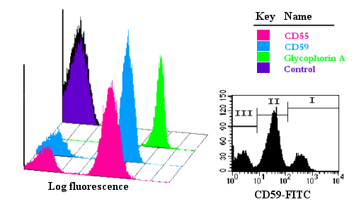
Physicians diagnose PNH using one of two blood tests — either a Ham test or flow cytometry. Treatments for PNH patients with hemolytic anemia include transfusion, folic acid, and if appropriate, iron supplements. Low red blood cell, platelet, and white cell counts can be treated with steroids or with the same immunosuppressive drugs used to treat aplastic anemia. Blood clots are usually treated with anticoagulants, and sometimes, if severe, with tissue plasminogen activator (TPA). PNH can be cured by bone marrow transplantation; this procedure should be considered on an individual basis, based on the patient’s age and symptoms.

TESTING FOR DIAGNOSIS OF PNH

Bone marrow samples can be obtained by aspiration and trephine biopsy. Sometimes, a bone marrow examination will include both an aspirate and a biopsy. The aspirate yields semi-liquid bone marrow, which can be examined by a pathologist under a light microscope as well as analyzed by flow cytometry, chromosome analysis, or polymerase chain reaction (PCR). Frequently, a trephine biopsy is also obtained, which yields a narrow, cylindrically shaped solid piece of bone marrow, 2mm wide and 2 cm long (80 μL), which is examined microscopically (sometimes with the aid of immunohistochemistry) for cellularity and infiltrative processes. An aspiration, using a 20 mL syringe, yields approximately 300 μL of bone marrow.[1] A volume greater than 300 μL is not recommended, since it may dilute the sample with peripheral blood.[1]


In a bone marrow analysis detection of the red cells will produce a defective protective layer. The white cell and platelets will be lower than normal . This result will decrease immunity in the body and therefore decrease resistance against disease. 

The Ham Test is also used in the diagnosis of PNH. 
HAM test is used to evaluate patients with suspected PNH (Paroxysmal Noctural Hemoglobinuria) or suspected congential dyserythropoietic anemia, especially with hemosiderinuria, Pancytopenia, decreased RBC acetyl cholinesterase, decreased leukocyte alkaline phosphatase, negative direct Coomb’s test, and/or apparent marrow failure.
Patients red cells show a high sensitivity to complement mediated hemolysis .

 LDH ( lactate dehydrgenase) is a blood test that is used to monitor tissue damage. The most important measure of hemolysis — can help provide a more complete clinical picture of PNH when performed in conjunction with other laboratory tests and patient-reported assessments. Monitoring of hemolysis in patients with PNH can be done by measuring LDH levels. LDH levels in PNH can be frequently elevated, in some cases exceeding 20 times the upper limit of normal during severe exacerbations.3 It is important to establish a baseline LDH level and continue monitoring over time. Static or “snapshot” measurements of LDH may not reflect the chronic and progressive course of the disease.


TREATMENT

Some of the supportive therapies that are currently being used to help ease the symptoms of PNH include transfusions and anticoagulants (Blood thinners)
Transfusions are recommended during times of severe haemoglobin depletion when your body cannot generate enough new cells to make up for those lost to haemolysis. It may be used on a periodic basis when your haemoglobin level is steadily decreasing.
Your physician may prescribe anticoagulants to you, in order to either reduce the risk of getting blood clots or may need to dissolve a clot when it happens.

Complement Inhibition:

When unregulated, the complement can lead to various severe diseases causing damages in multiple organs. Complement inhibitors are compounds which bind to enzymes in the complement system. Their role is to suppress or reduce the activity of complement.

Bone Marrow Transplantation:

Bone marrow transplantation (BMT) is the only known treatment that may cure PNH. It involves replacing the body’s defective blood stem cells by new healthy ones from a donor. However, bone marrow transplantations carry significant risks which should be discussed with your doctor.
It is important to remember that symptoms of PNH may vary from patient to patient, and a plan to manage your PNH may work for you but may not work for someone else. This is why it is so important to discuss treatment options with your doctor.


 The following websites are helpful.

http://www.pnhsource.eu/Diagnosing%20PNH/Flow%20Cytometry/Default.aspxhttp://www.mskcc.org/cancer-care/adult/rare-hematologic-disorders/paroxysmal-nocturnal-hemoglobinuria
http://www.medindia.net/bloodtest/hematology/pnh_test.htmhttp://pnhsource.eu/Treatment%20Options/Default.aspx

Sunday, December 4, 2011

Bone Marrow Biopsy

I wanted to educate about why a Bone Marrow is needed in diagnosing the reason of abnormal complete blood count results.  The Bone Marrow is the spongy tissue that is inside some of your larger bones such  as iliai crest which is the on the side of hip area.  Cells are produced in the bone marrow and should have normal amounts. If you have leukemia then there will be abnormal amount of cells.
In order to diagnose your medical condition the oncologist must do a Bone Marrow aspirate. Inside your bone is a fluid portion and a solid portion and this needs to be aspirated with a larger needle.


                                           Educational video of a Bone Marrow Aspirate


If your diagnosis is a blood cancer then you may have other bone marrow biopsies to see if your treatment is succeeding .  Your bone marrow will be put into several tubes and the core(bone part) will be put in formalin.
 The will perform genetic testing, flow cytometry , histology and stains.

  Complications to bone marrows are rare.  I would not take any aspirin and always make the doctor aware if you are taking any blood thinners.

Blood Disorders : Aplastic Anemia

Aplastic anemia is a condition where the bone marrow does not produce sufficient enough of new cells to replenish the blood cells in the body.


There are many circumstances why the bone marrow can shut down production of new cells.


  • Radiation and chemotherapy treatments. While these cancer-fighting therapies kill cancer cells, they can also damage healthy cells, including stem cells in bone marrow. Aplastic anemia can be a temporary side effect of these treatments.
  • Exposure to toxic chemicals. Exposure to toxic chemicals, such as some used in pesticides and insecticides, may cause aplastic anemia. Exposure to benzene — an ingredient in gasoline — also has been linked to aplastic anemia. This type of anemia sometimes gets better on its own if you avoid repeated exposure to the chemicals that caused your initial illness.
  • Use of certain drugs. Some medications, such as those used to treat rheumatoid arthritis and some antibiotics, can cause aplastic anemia.
  • Autoimmune disorders. An autoimmune disorder, in which your immune system begins attacking healthy cells, may involve stem cells in your bone marrow.
  • A viral infection. Viral infections that affect bone marrow may play a role in the development of aplastic anemia in some people. Viruses that have been linked to the development of aplastic anemia include hepatitis, Epstein-Barr, cytomegalovirus, parvovirus B19 and HIV.
  • Pregnancy. Aplastic anemia that occurs in pregnancy may be related to an autoimmune problem — your immune system may attack your bone marrow during pregnancy.
  • Unknown factors. In many cases, doctors aren't able to identify the cause of aplastic anemia. This is called idiopathic aplastic anemia.
Above is an image of Aplastic anemia bone marrow biopsy under a microscope.

How is Aplastic Anemia Diagnosed?
A complete blood count is performed and the cell counts, hemoglobin will be low.  Also a bone marrow biopsy will be performed and sent to pathology for the cytology technologists to stain and prepare to be read by the pathologist.  The report is then sent to your family doctor , specialist or oncologist.  Hematology oncologist is the doctor who should treat aplastic anemia. If you like to view a bone marrow biopsy then go tot the following website.http://youtube.com/watch?v=dTKAU34
Reticulocyte count is another test that is ordered in the diagnosis of Aplastic anemia. It is a test that measures how fast the bone marrow is producing new red cells. The reticulocyte test will be higher in if the hemoglobin is low and the bone marrow is trying to produce more cells due to blood loss.
Erythropoietin is a blood test that measures the amount of erythropoietin, a hormone which tells the bone marrow to produce more red cells.  This hormone is made by the cells in the kidney. The kidney produces more of the EPO hormone when oxygen levels are lower.  When someone is anemic they have trouble breathing because they do not have enough red cells to carry the oxygen.

Treatment for Aplastic anemia includes blood transfusions, platelet transfusions, a stem cell transplant, immunosuppressants, bone marrow stimulants and antiviral medications.

Anytime you may feel abnormally fatigued and have shortness of breath you need to see care from a medical professional.  You need to make sure that a complete blood count is performed .

http://www.mayoclinic.com/health/aplastic-anemia/; http://www.medline.com ; http://labsonline.com


Definition of Erythropoietin (EPO) Related Articles

Webster's New World
Medical Dictionary
Learn more »
Webster's New World Medical Dictionary MedTerms Medical Word
of the Day XML


<script language="JavaScript1.2" type="text/javascript" src="http://as.webmd.com/js.ng/Params.richmedia=yes&transactionID=466909359615&tile=466909359615&xpg=0&artid=7032&site=2&affiliate=22&uri=articlekey%3D7032&pos=113"></script>


Definition of Erythropoietin (EPO) Related Articles

Webster's New World
Medical Dictionary
Learn more »
Webster's New World Medical Dictionary MedTerms Medical Word
of the Day XML


<script language="JavaScript1.2" type="text/javascript" src="http://as.webmd.com/js.ng/Params.richmedia=yes&transactionID=466909359615&tile=466909359615&xpg=0&artid=7032&site=2&affiliate=22&uri=articlekey%3D7032&pos=113"></script>


Friday, December 2, 2011

Blood Disorders: Polycythemia Vera

Polycythemia vera is not a cancer,but is a disease of the bone marrow.  Hematology oncologists handle the diagnosis and treatment of this disease. The bone marrow makes too many red cells.  Polycythemia is rare and develops slowly.  The problem of elevated red blood cells is usually found in a routine complete blood count which is ordered by doctor.   The image below shows what polycythemia vera looks like under the microscope.


If  polycythemia vera is not treated then it could become life threatening. The symptoms are the following:
In its early stages, polycythemia vera usually doesn't cause any signs or symptoms. However, as the disease progresses, you may experience:
  • Headache
  • Dizziness
  • Itchiness, especially following a warm bath or shower
  • Redness of your skin
  • Shortness of breath
  • Breathing difficulty when you lie down
  • Numbness, tingling, burning or weakness in your hands, feet, arms or legs
  • A feeling of fullness or bloating in your left upper abdomen due to an enlarged spleen
  • Fatigue  
Blood tests
Doctors most frequently use blood tests to diagnose polycythemia vera. If you have polycythemia vera, blood tests may reveal:
  • An increase in the number of red blood cells and, in some cases, an increase in platelets or white blood cells.
  • Elevated hematocrit measurement, the percentage of red blood cells that make up total blood volume.
  • Elevated levels of hemoglobin, the iron-rich protein in red blood cells that carries oxygen.
  • Very low levels of erythropoietin (EPO), a hormone that stimulates bone marrow to produce new red blood cells.
Bone marrow aspiration or biopsy
If your doctor suspects you have polycythemia vera, he or she may recommend a bone marrow aspiration or biopsy to collect a sample of your bone marrow. A bone marrow biopsy involves taking a sample of solid bone marrow material. A bone marrow aspiration is usually done at the same time as a biopsy. During an aspiration, your doctor withdraws a sample of the liquid portion of your marrow.
If an examination of your bone marrow shows that it's producing higher than normal numbers of blood cells, it may be a sign of polycythemia vera.
Tests for the gene mutation that causes polycythemia vera
If you have polycythemia vera, analysis of your bone marrow or blood also may show the mutation in the cells (JAK2 V617F mutation) that's associated with the disease.
n a bone marrow aspiration and biopsy, a doctor or nurse uses a thin needle to remove a small amount of liquid bone marrow, usually from a spot in the back of your hipbone called the posterior iliac crest. A bone marrow biopsy is often taken at the same time. This second procedure removes a small piece of bone tissue and the enclosed marrow.

Treatment of polycythemia include doing a phlebotomy to remove blood that is putting your circulatory system in overload.  It is similar to giving blood to the Red Cross.  Drugs are given if the phlebotomy is not enough to relieve the increased red cell production.  
 Hydroxyurea (Droxia, Hydrea) or anagrelide (Agrylin), to suppress your bone marrow's ability to produce blood cells may be used. Interferon-alpha may be used to stimulate your immune system to fight the overproduction of red blood cells.
Any abnormal symptoms should always be addressed by a doctor.  Be in tune with your body and document changes to help the doctor to have a clearer picture of your health problems.

You can get more information be searching https:www.mayoclinic.com