Saturday, May 12, 2012

Sharing Article of Breast Cancer Diagnostic Tool

This is great news that was published this past week and it must be shared with all women in the fight against breast cancer. Please share this article with a friend .

Below is an image of detecting chromosome translocations in sections of breast. 
 In breast cancer, we have shown that the NRG1/heregulin gene is translocated in 6% of primary cases and Soda and colleagues described fusions of ALK in 7% of lung cancers .
 

 Article

Pathologists today know that there are certain gene mutations that are predictive of cancers; for example, women with the BRCA1 or BRCA2 mutation are at a greater risk of developing breast and/or ovarian cancer. What is less known is the availability of genetic HER-2/neu FISH testing, a sophisticated laboratory test able to better determine treatment options for an aggressive form of breast cancer. This information is important because it helps doctors to determine the best course of treatment for each patient.
“HER-2/neu FISH testing is considered the gold standard because it has the advantage of looking at each tumor cell individually,” said Olga Falkowski, MD, a board-certified pathologist who serves as the Associate Medical Director and Director of Genetics at Acupath Laboratories, Inc., a national medical laboratory based in Plainview, NY. “Under U.S. Food & Drug Administration guidelines, all women with invasive breast cancer are eligible to undergo the test.”
In the body, the HER protein is a normally occurring substance that helps cells grow and stay strong. In about 20 percent of breast cancers, patients are HER-2 positive, meaning that cancer cells underwent genetic mutations responsible for production of excess protein called the HER-2/neu or human epidermal growth factor receptor 2. Patients with the genetic mutation HER-2 in breast cancer are eligible for a targeted therapy. Because of this, both the American Society for Clinical Oncology and National Comprehensive Cancer Network recommend HER-2/neu testing for all breast cancer tumors.
Women who undergo a biopsy for a suspicious lump and are diagnosed with cancer first receive immunohistochemistry or IHC of HER-2/neu testing as part of their initial laboratory workup. If the IHC testing is positive for Her-2/neu, then FISH is performed to confirm the genetic mutation.
What makes FISH or fluorescence in situ hybridization testing different and generally more effective, Dr. Falkowski said, is that it examines each individual cell of the tumor. “That’s a huge advantage,” she added. “The more a doctor knows about a tumor and its characteristics, the better that doctor can determine the most effective treatment options. The HER-2/neu FISH test tends to be more reliable.”
IHC testing quality can vary between laboratories based on the type of antibodies the lab uses, the way the lab prepares the tissue sample, and its criteria for determining the presence of HER-2, which is not the same everywhere. “Different labs have different reading systems,” Dr. Falkowski explained.
FISH testing is a more complex procedure, but is more specific and sensitive. It uses fluorescent probes to identify, tag and count the presence of genes that cause excessive production of the HER-2 protein in each individual cell. If more than two copies of the gene are found in each cell, the cancer is determined to be HER-2 positive and treated as such.
“It’s a more reproducible test, meaning the results from one lab to another should be the same,” Dr. Falkowski explained, adding that the HER-2 protein can also affect the aggressiveness and treatment of gastric and ovarian cancer. “Because it’s expensive technology, HER-2/neu FISH testing isn’t available everywhere, so doctors and their patients should know to ask for it,” adds Dr. Falkowski.
A concrete, reliable diagnosis of whether a woman with breast cancer is HER-2 positive will help lead to the best possible treatment starting immediately, improving survival and helping prevent recurrence. Standard chemotherapy drugs can be effective in treating Her-2/neu-positive patients, though more effective are two drugs that specifically target the Her-2/neu protein, trastuzumab (Herceptin) and lapatinib (Tykerb). Trastuzumab can be used alone or in conjunction with chemotherapy or hormone-blocking medications, though it comes with some potentially risky side effects, including congestive heart failure. Lapatinib is also generally used in combination with chemotherapy, most often in patients who have problems with trastuzumab. A promising new drug called trastuzumab emtansine or T-DM1 that combines several agents is also currently in clinical trials.
Dr. Falkowski continued, “As dramatic as this may sound, it’s true: For some women, HER-2/neu FISH testing could mean the difference between life and death.”
Olga Falkowski, M.D. is board-certified in anatomic and clinical pathology by the American Board of Pathology, and serves as the Associate Medical Director and Director of Genetics at Acupath Laboratories, Inc.
Acupath Laboratories, Inc. is a Plainview, New York, specialty medical lab engaged in cutting-edge diagnostics. http://www.acupath.com

http://www.prweb.com/releases/2012/5/prweb9457999.htm

Friday, May 11, 2012

Metastatic Breast Cancer

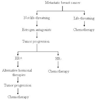
Metastatic breast cancer is a stage of breast cancer where the disease has spread to distant metastases. It is a complication of primary breast cancer, usually occurring several years after resection of the primary breast cancer. Metastatic breast cancer cells frequently differ from the preceding primary breast cancer in properties such as receptor status, have often developed resistance to several lines of previous treatment and acquired special properties that permit them metastasize to distant sites, making them especially dangerous. The prognosis is often poor, distant metastases are the cause of about 90% of deaths due to breast cancer.

Breast cancer primarily metastasizes to the bone, lungs, regional lymph nodes, liver and brain, with the most common site being the bone. Lymph node metastsasis into the sentinel node and few surrounding nodes is regarded as a treatable local event and not metastatic breast cancer, both when occurring at primary presentation or later.


                                                Progression of Metastatic Breast Cancer


                       Advancements in treating metastatic cancer by Professor Steven Twelves M.D.

                                         Faces of those with Metastatic Cancer


Today, say the MD Anderson researchers, as many as 40% of women with recurrent or metastatic breast cancer survive at least five years. "More and more, both doctors and patients approach it as a chronic condition," says Eric Winer, MD, director of the Breast Program at Boston's Dana-Farber Cancer Institute. "We can't cure it, but we can manage it for many years."

For more information:http://www.webmd.com/breast-cancer/features/metastatic-breast-cancer-chronic-condition

Thursday, May 10, 2012

Merkel Cell Carcinoma

Merkel cell carcinoma is a rare and aggressive cancer. It is usually found in older patients and a compromised immune system. There are about 1500 reported cases per year but the number is increasing. One -third of patients could die quickly from being diagnosed with MCC and need to be treated immediately.

MCC is also referred as neuroendocrine carcinoma and grows in large amounts in the skin. Over weeks a small bump grows rapidly.


It is very difficult to look at the horrible picture,but this MCC growth can be seen on any part of the body.

This is a picture of Merkel Cell Carcinoma under a microscan which magnifies very high.

Merkel cells are found in the epidermis (outer layer of the skin). Although the exact function of Merkel cells is unknown, they are thought to be touch receptors. They have both sensory and hormonal functions and are sometimes referred to as neuroendocrine cells.

Dr. Randall K. Roenigk of the prestigious Mayo Clinic explains Merkel Cell carcinoma.

What is the typical patient like that can get MCC?  They are usually 65 or over,fair skinned,experienced lots of exposure to the sun and immuno depressed. Also persons who are HIV positive may be susceptible because this disease compromises their immune system.

The following testing is performed to diagnose MMC:

  • Sentinel node biopsy. When cancer cells spread, they often travel first to your lymph nodes — small, rounded structures that filter foreign particles from lymph, a tissue-cleansing fluid in your body. A sentinel lymph node biopsy is a procedure to determine whether cancer has spread to your lymph nodes. This procedure involves injecting a dye near the skin tumor. The dye then flows through the lymphatic system to your lymph nodes. The first lymph node that receives the dye is called the sentinel node. Your doctor removes this lymph node and looks for cancerous cells under a microscope.
  • Imaging tests. Your doctor may recommend a chest X-ray and a CT scan of your chest and abdomen to help determine whether the cancer has spread to other organs. Your doctor may also consider other imaging tests such as a positron emission tomography (PET) scan or an octreotide scan — a test that uses an injection of a radioactive tracer to check for the spread of cancer cells.
Treatment for MCC  is first of surgery to attempt to remove the carcinoma. Radiation is the next step to reduce the growth of remaining cancer cells. Finally, chemotherapy is used to also decrease continued growth and kill the growing tumor.

The following website is a wonderful place to view for information and support for anyone with MCC.
http://www.merkelcell.org/faqs/index.php#aboutDisease_01


Wednesday, May 9, 2012

Protocol In Place For Pediatric Cancer Patients Arrival to E.R.

Parents of children with cancer will be pleased that pre-arrival protocol has been placed in some Emergency Departments to shorten the time for the pediatric patient. Children with cancer can easily have fevers that need to be addressed due to a compromised immune system because of chemotherapy. Parents are already very stressed due to what it is required dealing with all the scheduled treatments at cancer centers. They do not need to have deal with a long wait when they sense that their child is seriously ill.

Recent news of a plans in place for every pediatric patient diagnosed with cancer.
By implementing a standardized referral notification to the ED physician and charge nurse, using a standardized electronic health-record-based referral checklist with patient-specific information, and using a standardized order set for laboratory tests and antibiotics at the time of referral, investigators reduced the median time to antibiotic administration by almost an hour, according to Evaline Alessandrini, MD, MSCE, from the division of emergency medicine at the Cincinnati Children's Hospital Medical Center and professor of pediatrics at the University of Cincinnati College of Medicine in Ohio.
Dr. Evaline Alessandrini
Dr. Alessandrini explained that the timely delivery of antibiotics to febrile children with cancer presented an opportunity to test elements of the Chronic Care Model related to health systems and to the organization of healthcare. Previsit planning is a critical part of chronic disease management. Specifically, delivery system design, decision support, clinical information systems, and a prepared practice team lend themselves to the development of a prearrival plan.
Our data demonstrated that the largest gaps in care are between ED arrival and the time that orders for antibiotics are written, Dr. Alessandrini explained. "We planned to close the gap by ordering the antibiotics before the patient arrived in the ED."
For trauma alerts, roles are assigned and equipment and medications prepared before the patient arrives. "Our goal was to transfer these activities to the ED during the management of acute exacerbations of chronic disease," she said.

The researchers wanted to reduce the time from patient arrival to the administration of antibiotics for pediatric cancer and bone marrow transplantation patients with fever from 140 minutes to less than 90 minutes. The setting was an urban tertiary-care children's hospital ED. The hospital has a high-volume oncology and bone marrow transplantation service that sees febrile patients in its clinic during weekday daytime hours. At other times, the children are seen in the ED. Because all of the children are immunosuppressed, there is no reason to wait for a complete blood count before giving antibiotics.

This plan has reduced time in the Emergency Department for about 100 minutes.


For more info: http://www.medscape.com/viewarticle/763467

Tuesday, May 8, 2012

New Cervical Cancer Screening to Hit World Wide Market

This new cervical cancer screening still involves a pap smear,but it reduces the number of false positives in the result of testing. There is also other changes that make the screening more cost effective.

Biodis is the name of this biotechnology distributor that will launch their new product to many countries of the world. Citofem is the name of the actual test.

"There are only two major pap smear products on the market today, now with Citofem(R), there is a third. This Pap test kit however, offers the highest sensitivity at the lowest price, making this a powerful weapon in the battle against cervical cancer worldwide," said Kent Erickson, Vice President of Sales for North America.

Why is this screening pap smear product better than the other two on the market? Citofem give the same superior results as the other two previous tests but it allows for a manual procedure that is very easy to replaces the purchase of expensive lab equipment.The process is extremely efficient with a single cytotechnologist processing on average over 750 slides per day.

Citofem results in a significant reduction in false negatives, increasing the diagnosis of the disease in its early stages," said Dr. Ladly Abraham, Chief Medical Officer of Biodis. "Citofem is also responsible for a reduction in inconclusive results arising from atypical squamous cells (ASC). It also results in the increased level of detection of low grade squamous intraepithelial lesions (LSIL) and high-grade squamous intraepithelial lesions (HSIL). We're very excited about Citofem because it's the most complete system for the early detection of cervical cancer, in that it obtains a much higher population of endocervical cells in the sample."

Top Honors For the Outstanding Innovation of the development of  Citofem Pap Smear Screening.

For more info:
http://www.marketwatch.com/story/revolutionary-new-cervical-cancer-screening-test-to-hit-worldwide-market-2012-05-02

Thursday, May 3, 2012

Quest Diagnostics Show Support For the Cancer Testing

A new effort in urging women to be proactive in cancer prevention.  The American Cancer society is promoting the "Choose You® movement for women's health. Quest Diagnostics has decided to be a large part of this undertaking. The following information was released in the news.


Quest Diagnostics (NYSE: DGX), the world's leading provider of diagnostic testing, information and services, today announced its national presenting sponsorship of the American Cancer Society Choose You® movement, which encourages women nationwide to live well today and stay well tomorrow. In support of the Choose You movement and in recognition of National Women's Health Week May 13 – 19 sponsored by the U.S. Department of Health and Human Services Office on Women's Health, Quest Diagnostics will offer 10,000 vouchers for free screenings to women at May Choose You events around the country, beginning today in New York City.
"One in three women will get cancer in her lifetime. But a significant number of cancer deaths could be prevented if people maintained a healthy weight through diet and regular exercise, avoided tobacco products and had their regular health checks," said Kimberly Wright, Director, Mission Solutions and Tools, at the American Cancer Society. "We're proud to have Quest Diagnostics as our Choose You national presenting sponsor, and we greatly value their partnership as we work together to empower women to get active, manage their health, and stay well."
An American Cancer Society survey finds that 40 percent of women said they would be more physically active in their free time if it felt less like work and more like play. In response, Choose You is issuing a challenge to women: Choose play in May and help the Society inspire 100,000 acts of physical activity. To get the challenge started, a series of Choose You pop-up "play teams" will be hitting the streets of New York (May 1), Los Angeles (May 11) and Washington, DC (May 22), inviting passers-by to get active with nostalgic childhood toys, including the '80s famed Skip It, hula hoops, hoppity-hop balls and double Dutch jump ropes. Today the Choose You team launched the movement by encouraging women to play midday through late afternoon in New York's Times Square.
"Choose You is an important and growing women's health initiative, and it made great sense in 2012 for Quest Diagnostics to continue and strengthen our support of the women touched by this American Cancer Society movement," said Laure Park, Quest Diagnostics' Corporate Citizenship Officer. "Whether you're a family member, healthcare provider, co-worker or friend, we hope you'll join us – this month and throughout the next year – in encouraging the women in our lives to prioritize a healthy lifestyle, get out and play, and make time for their regular health checks.

Read more here: http://www.sacbee.com/2012/05/01/4457562/quest-diagnostics-sponsors-american.html#storylink=cpy

Wednesday, May 2, 2012

Facts about Ovarian Metastastis From the Colon

According to the Mayo Clinic, "Symptoms of ovarian cancer are nonspecific and mimic those of many other more common conditions, including digestive and bladder disorders." Colon cancer may also spread to the lymph nodes, bones, lungs and liver. There are particular signs and symptoms that the cancer has spread to the ovaries.

Abdominal Discomfort

  • Patients with colon cancer that has spread to the ovaries may have already been experiencing pain, fullness or bloating in the abdomen from the colon cancer and may not realize the cancer has spread. Patients may also experience persistent indigestion, gas or nausea as well as changes in bowel habits, such as constipation. One may lose their appetite or quickly feel full after a meal.

Pelvic Discomfort

  • Women may experience pain during intercourse, lower back pain and general pelvic discomfort. According to the Mayo Clinic, patients may also experience changes in bladder habits, including a frequent need to urinate as well as changes in menstruation (more bleeding, much less bleeding or erratic bleeding).
The following is an abstract from a Colorectal Medical Journal

Abstract

Objective  To improve management of ovarian metastasis through assessment of clinicopathological features and treatment outcomes associated with ovarian metastasis from colorectal cancer.
Method  We recruited 103 subjects who were diagnosed with ovarian metastasis and subjected to surgery between June 1989 and December 2005. Clinical and pathological variables were evaluated. Survival and its associated factors were analysed with a median follow-up of 31 months after ovarian surgery (range 1–129 months).
Results  The mean age at diagnosis was 46 years (range 14–72 years), synchronous ovarian metastasis occurred in 74 patients and metachronous in 29 patients. The primary tumour was more commonly associated with the colon rather than the rectum (84/1608, 5.2%vs 19/1534, 1.2%, < 0.001). Combined metastases occurred in 69 patients (67%). Complete resection was achieved in 34 (33%) patients without other metastases. The estimated 5-year disease free survival and overall survival rate were 40.1% and 26.6%, respectively. From univariate analysis, lymphovascular invasion (35.6%vs 12.8%, = 0.034), combined metastasis (50.9%vs 15.6%, = 0.0035) and bilaterale ovarian metastasis (36.4%vs 10.6%, = 0.015) were identified as significant poor prognosis factors, and from multivariate analysis combined metastasis and bilaterale ovarian metastasis were significant (= 0.034 and = 0.015, respectively).
Conclusion  This study suggests a role for regular follow-up computed tomography scans within 6 months postoperatively and tumour marker assays for the early detection of ovarian metastasis in premenopausal women after primary surgery, especially in colonic patients with poor prognostic factors.

Diagnosis

A pelvic examination and imaging including CT scan[39] and trans-vaginal ultrasound are essential. Physical examination may reveal increased abdominal girth and/or ascites (fluid within the abdominal cavity). Pelvic examination may reveal an ovarian or abdominal mass. The pelvic examination can include a Rectovaginal component for better palpation of the ovaries. For very young patients, magnetic resonance imaging may be preferred to rectal and vaginal examination.
To definitively diagnose ovarian cancer, a surgical procedure to take a look into the abdomen is required. This can be an open procedure (laparotomy, incision through the abdominal wall) or keyhole surgery (laparoscopy). During this procedure, suspicious areas will be removed and sent for microscopic analysis. Fluid from the abdominal cavity can also be analysed for cancerous cells. If there is cancer, this procedure can also determine its spread (which is a form of tumor staging).

Staging

Ovarian cancer staging is by the FIGO staging system and uses information obtained after surgery, which can include a total abdominal hysterectomy, removal of (usually) both ovaries and fallopian tubes, (usually) the omentum, and pelvic (peritoneal) washings for cytopathology. The AJCC stage is the same as the FIGO stage. The AJCC staging system describes the extent of the primary Tumor (T), the absence or presence of metastasis to nearby lymph Nodes (N), and the absence or presence of distant Metastasis (M).[43]
  • Stage I — limited to one or both ovaries
    • IA — involves one ovary; capsule intact; no tumor on ovarian surface; no malignant cells in ascites or peritoneal washings
    • IB — involves both ovaries; capsule intact; no tumor on ovarian surface; negative washings
    • IC — tumor limited to ovaries with any of the following: capsule ruptured, tumor on ovarian surface, positive washings
  • Stage II — pelvic extension or implants
    • IIA — extension or implants onto uterus or fallopian tube; negative washings
    • IIB — extension or implants onto other pelvic structures; negative washings
    • IIC — pelvic extension or implants with positive peritoneal washings


 Advanced Ovarian Cancer
Ovarian adenocarcinoma deposit in the mesentry of the small bowel
  • Stage III — peritoneal implants outside of the pelvis; or limited to the pelvis with extension to the small bowel or omentum
    • IIIA — microscopic peritoneal metastases beyond pelvis
    • IIIB — macroscopic peritoneal metastases beyond pelvis less than 2 cm in size
    • IIIC — peritoneal metastases beyond pelvis > 2 cm or lymph node metastases
  • Stage IV — distant metastases to the liver or outside the peritoneal cavity
                                   Testimonial of Patient with Colon Cancer that Metastasized to Ovary

http://ovariancancer.jhmi.edu/prognosis.cfm

http://www.ncbi.nlm.nih.gov/pmc/articles/PMC2650975/