Saturday, December 29, 2012

Research In Using Boron As a Anti-Cancer Agent Is Recognized


Would it not be fantastic to be able to reduce radiation cancer treatment to one session. Those who have or had cancer may have experienced several treatments of radiation and they can really take its toll on the human body.


Fred Hawthorne who wants to kill cancer with tiny nuclear bombs loaded into each diseased cell will be honored early next year at a White House ceremony celebrating the nation's top scientists and innovators.

Hawthorne is pioneering the use of boron as an anti-cancer agent. His research, he said yesterday, is intended to replace lengthy and painful radiation treatments with one effective session that wipes out the diseased tissue.

To do so, he and his team are developing techniques that load the cancer cells with boron. The tissue is then bombarded with neutrons, which are easily absorbed by the boron.

"That capture event causes a tiny nuclear explosion, which degrades the boron atom and the neutron," Hawthorne said. "That releases a lot of energy locally, and it kills it very selectively."

"We have everything we need provided by the university, and we are very, very thankful," Hawthorne said. "I feel they have done very, very well by us, and we are bringing home the bacon."

A set of animal trials testing the use of boron as an anti-disease agent went well, and Hawthorne said the research is being prepared for publication. He expects the treatments might be ready for human use in about five years.

"There is no reason it shouldn't work, given a chance," he said. "What we needed is a demonstration in animals that it is effective, and that is coming along very nicely."

Boron capture therapy also holds promise as a potential treatment for arthritis, heart disease and Alzheimer's, an MU news release said. "When Dr. Hawthorne came to UM in 2006, I was sure that he would advance MU's national leadership in nanomedicine and cancer research while providing breakthrough technology and medical solutions for the world," Chancellor Brady Deaton said in the news release. "This acknowledgement by President Obama of Dr. Hawthorne's work is especially gratifying and well deserved."

Hawthorne, 84, has more than a theoretical interest in the research. Since arriving in Columbia, he has been treated for a cancerous growth on his tongue, which required surgery, a dozen chemotherapy treatments and 35 radiation treatments. If boron treatment had been available, it would have replaced the radiation therapy, he said.

The boron is delivered in a method that takes it directly into the cancer cell and no other cells of the body, he said. "It goes to the proper place, it is irradiated down there and it's finished. It is all over, and that is all he needs."


Monday, December 10, 2012

New Pancreatic Cancer Research Laboratory Opened

The Lustgarten Foundation and Cold Spring Harbor Laboratory (CSHL) report that the new Lustgarten Foundation Pancreatic Cancer Research Laboratory is now in operation. The lab’s initial studies will center on early detection, drug development, and drug deliver.


Pancreatic cancer is the most deadly of all cancers. Early detection and new drug therapy is the key to aggressively destroy cancer cells in this vital organ in out bodies.

In the medical testing community there is no test that can detect early stages of pancreatic cancer and no cancer drug therapy that can completely destroy the cancer cells in the pancreas.


The Tuveson lab has developed mouse models of pancreatic ductal adenocarcinoma (PDAC) to discover biomarkers of early disease and to identify the pathways and druggable targets involved in the initiation, progression, and metastasis of PDA. Efforts are also underway to come up with efficacious therapeutic strategies.

“We have discovered that PDAC tumors contain a deficient and compressed vasculature that limits therapeutic delivery and therefore efficacy,” notes Dr. Tuveson. “Using these models we have uncovered several methods to correct or target these vascular deficits and promote response, and this information has led to the initiation of several clinical trials.

“At CSHL we will search for new vulnerabilities in PDAC neoplastic and microenvironmental cells, and evaluate these candidates in a futuristic 'Mouse Hospital' we are creating on campus.”


Wednesday, November 28, 2012

Two Bio-science Companies United to Help in the Diagnosis of Lung Cancer


Bioview will develop in automated cancer screening system to carry out Abbott Molecular's lung cancer diagnostic test.


Cancer screening company BioView Ltd. (TASE:BIOV) has signed a cooperation agreement with Abbott Molecular Inc., under which Bioview will develop in automated cancer screening system to carry out Abbott's lung cancer diagnostic test.

Abbott Molecular's current test to distinguish between different kinds of lung cancer, in order to provide the proper treatment, is carried out manually by a laboratory technician. Automating the test will save time and effort, and improve accuracy.

BioView has already applied its technology to other Abbott Molecular tests for breast and bladder cancer. Each time that these tests were approved, they boosted BioView's sales by millions of dollars.

Under the new collaboration, the two companies will jointly conduct clinical trials of the automated lung cancer test in order to certify it for marketing in the US. Abbott's manual test is already certified by the US Food and Drug Administration (FDA). The companies estimate that this process will take several months.

Lung Cancer: Molecular Tumor Testing Video


Wednesday, November 14, 2012

New York Department of Health Guidelines for Assist Labs in Cancer Testing


You may be wondering what type of guidelines has been developed by the New York Department of Health for labs. It is in the developing NGS based cancer assays. NGS stands for Next Generation Sequencing.

Of all the many topics under discussion at the NCI Clinical NGS for Cancer conference, perhaps the most contentious was the issue of which tests should be performed and how they should be reported. In this instance, the great advantage of the new technology—inexpensive and high-volume sequence data—is also in some ways its Achilles heel.


The guidelines, which cover somatic variant detection for molecular oncology testing, were issued in August by the department's Clinical Laboratory Evaluation Program, or CLEP, which monitors the quality of testing conducted by clinical laboratories and blood banks in the state as well as out-of-state labs that accept clinical samples from New York.

The document complements existing department guidelines for molecular oncology tests, following "the same basic principles for validating most other complex molecular diagnostics procedures," and are expected to evolve "as the field matures and gains experience," according to the document.


Erasmus Schneider, director of CLEP's oncology section at the department's Wadsworth Center in Albany, told Clinical Sequencing News that the need for the guidelines arose because next-gen sequencing is making its way into clinical diagnostics and the department had received requests from laboratories wanting to know how to validate their tests in order to gain New York State approval.

Input for the guidelines came from meetings Schneider and his colleagues attended where the clinical use of NGS was discussed and from feedback from interested parties on a draft document.

NGS-based molecular oncology testing differs from most other tests in this area because it interrogates panels of tens to hundreds of mutations in parallel rather than individually. Schneider said the only other high-throughput genotyping assay currently used for oncology mutation detection is the Sequenom MassArray assay, but that this "has not been widely adopted" by clinical laboratories in the state, in part because of the big investment required. "I think most people are much more familiar with sequencing-based assays," he said.


So far, the majority of clinical laboratories offering somatic mutation detection for cancer in New York have expressed an interest in NGS-based assays, Schneider said. Overall, 104 labs in the state hold a permit for molecular oncology testing, but not all of them offer somatic mutation detection, he added.

No NGS-based oncology tests have been approved yet, but several applications are pending.

Cyrus Hedvat, director of the diagnostic molecular pathology lab at Memorial Sloan-Kettering Cancer Center, said that the guidelines provide "a solid starting point for labs in the process of developing clinical next-generation sequencing-based tests."

His lab is currently developing an assay for solid tumors that runs on Illumina's MiSeq and uses the firm's TruSeq Amplicon Cancer Panel, which includes 212 amplicons from 48 cancer genes (CSN 10/17/2012).

Among other requirements, New York State's guidelines call for a laboratory to define the minimum coverage required for calling a variant with high confidence and for calling an amplicon normal. According to Hedvat, this is similar to guidelines published by the College of American Pathologists this summer (CSN 8/1/2012), but New York offers "no specific guidance on the coverage depth required or the minimum allelic frequency to report a positive result."


"This will be a significant advantage for common mutations in genes with hotspots such as EGFR, KRAS, and BRAF, but more problematic for a tumor suppressor such as P53," Hedvat said.

He also said that the guidelines are unclear about how many positive controls need to be included in each run, and whether they have to cover mutations from all regions tested. The guidelines do state that a positive control has to be included during validation "and periodically thereafter," and suggest using a control containing "multiple known somatic alterations of each kind to be detected."

With regard to reports, the guidelines state that they should include all detected somatic variants, even those of unknown significance. "This could result in a long list of variants that could be difficult for a clinician to interpret," Hedvat said, but on the other hand, if later publications make such variants clinically interpretable, the lab would not need to issue an updated report.

                                       Next Generation Sequencing Technologies Explained


Monday, October 29, 2012

Antidepressant Used to Ease Pain During Oral Cancer Treatment


Doxepin, an antidepressant used to treat a combination of symptoms of anxiety and depression, can also significantly ease pain associated with oral mucositis in patients receiving radiation therapy for cancers of the head and neck, according to a Mayo Clinic study presented today at the American Society for Radiation Oncology annual meeting in Boston.

Oral mucositis is the painful inflammation and ulceration of the mucous membranes lining the mouth, gums, tongue, and throat. It is a common and often debilitating side effect of radiation therapy and chemotherapy cancer treatments. But ongoing research into the effects of doxepin on oral mucositis shows that the pain associated with this kind of inflammation can be significantly reduced.

Doxepin is sold under the brand names Adapin, Silenor, and Sinequan, among others.


In this study, researchers found that doxepin was well-tolerated and eased pain among patients with oral mucositis. However, a few subjects reported side effects, such as stinging, burning, unpleasant taste, and drowsiness. Given the option, 64 percent of the 155 participants decided to continue using doxepin after the study was complete.

"Oral mucositis or mouth sores is a painful and debilitating side effect of radiation therapy," said principal investigator Robert Miller, M.D., a radiation oncologist at Mayo Clinic. "Our findings represent a new standard of care for treating this condition."

While participants reported a few side effects, Miller said doxepin rinse does not cause the side effects associated with narcotic pain medicines, making it a better treatment for oral mucositis.


                                                            Cancer Facts: Oral Cancer

Monday, October 15, 2012

Hope in a Cancer Vaccine for Cervical Cancer

Cervical cancer is cancer that starts in the cervix, the lower part of the uterus (womb) that opens at the top of the vagina.  Worldwide, cervical cancer is the third most common type of cancer in women. It is much less common in the United States because of the routine use of pap smears

Cervical cancers start in the cells on the surface of the cervix. There are two types of cells on the cervix's surface: squamous and columnar. Most cervical cancers are from squamous cells.

ERIE TIMES-NEWS 

A vaccine against cervical cancer, being developed by Inovio Pharmaceuticals Inc. of Blue Bell, produced positive results in a small sample of 18 women.


The vaccine prompted their bodies to produce T cells -- a type of white blood cells -- that, in a separate lab test, recognized cells with tumor proteins, and killed them.


The researchers, including a team from the University of Pennsylvania, say the paper in the journal Science is the first to show that a DNA vaccine alone produced a high level of immunity in people. At the same time, the researchers acknowledged that a working vaccine faces more trials and remains years away from an actual product.

                                  What is Cervical Cancer Video 2012


Monday, October 8, 2012

New Biomarkers Have Been Developed to Help Monitor Cancer Treaments

Biomarkers are used in helping oncologists to diagnose several types of cancer. They are also used to monitor chemotherapy and radiation treatment in order for the doctor to know if they are killing the cancer cells. New biomarkers have been developed to provide more options in helping the different areas of oncology to treat patients.    


http://www.bizjournals.com/stlouis/blog/BizNext/2012/10/lee-biosolutions-expands-cancer-marker.html


Lee Biosolutions Inc. has expanded its line of biomarkers to include products used to track epithelial ovarian cancer, breast cancer and liver cancer.

Based in Brentwood, Lee Biosolutions collects biological materials — human saliva, semen, urine and vaginal secretions, among others — and produces finished proteins, enzymes, biologicals, immuno-reagents and antibodies for life science research and clinical diagnostic testing. The company’s core business is diagnostic and Lee Biosolutions sells proteins and enzymes to research labs like the Cleveland Clinic and pharmaceutical companies such as Abbott Laboratories.

Lee Biosolutions has developed a proprietary process to purify and test for novel cancer biomarkers. In addition to the new biomarkers, the company is expanding its production of the other biomarkers in its lineup.

Earlier this year, the company said it had developed a unique process for purifying the prostate cancer tumor marker PSA.

"We continue to increase supply of CA 19-9 which often is used with patients with pancreatic cancer, CEA tumor marker that helps predict the outlook in patients with colorectal cancer and CA 72-4 which is now used in ovarian, pancreatic cancer and cancers starting in the digestive tract," said Burton Lee, president of the company. "Current research has identified CYFRA 21-1 used as a marker for non-small cell lung cancer and Lee Biosolutions has increased supply of Beta 2 Microglobuilnwhich is used as a marker for multiple myelomas and chronic lymphocytic leukemia."

The products are raw finished proteins that are used in formulations in invitro diagnostic products and research.

Lee Biosolutions had revenue of $6 million last year.                                         

Monday, September 24, 2012

New Drug for Soft Tissue Sarcoma Doing Well in Clinical Trials

Sarcoma is cancer of the connective tissue such as muscle, cartilage, blood vessels, nerves and bones. Soft tissue (non-bone) sarcomas are rare, but they can occur in many parts of the body such as muscle or fat of the extremities or the trunk. They can also occur in the abdomen, pelvis or chest. There are many types of soft tissue sarcomas, but most are treated in the same way. Some common soft tissue sarcomas include fibrosarcoma, liposarcoma, malignant fibrous histiocytoma, rhabdomyosarcoma and synovial sarcoma.


CytRx Corporation a biopharmaceutical company specializing in oncology, today announced that favorable results from a Phase 1b/2 clinical trial with aldoxorubicin (formerly INNO-206) in patients with advanced soft tissue sarcoma will be featured in a poster and discussion presentation at the Connective Tissue Oncology Society (CTOS) 17th Annual Meeting on November 15 in Prague, Czech Republic.

The abstract, "Aldoxorubicin (INNO-206) is an active drug for the treatment of relapsed or refractory soft tissue sarcomas," will be presented by the clinical trial's principal investigator Dr. Sant Chawla, M.D., F.R.A.C.P. Dr. Chawla, a world renowned expert in the treatment of soft tissue sarcomas, is Director of the Sarcoma Oncology Center in Santa Monica, Calif. The Phase 1b/2 clinical trial results, first announced in early June 2012 at the American Society of Clinical Oncology (ASCO) conference, showed clinical benefit (defined as partial response and stable disease of more than four months following up to eight cycles of treatment) with aldoxorubicin at the maximum tolerated dose in 10 of 13 evaluable patients with advanced, metastatic soft tissue sarcomas who had either not responded or relapsed after receiving one to three prior chemotherapies. Eight of the 13 patients had been treated previously with doxorubicin.

"We are excited to share the powerful data from our aldoxorubicin clinical trial with experts from the international oncology community at this preeminent sarcoma conference, as well as at the upcoming ESMO 2012 Congress in Vienna, Austria," said Steven A. Kriegsman, CytRx President and CEO. "The results from this clinical trial are impressive, especially given that these patients had failed prior chemotherapy regimens. Further, there were no observed cardiac toxicities with aldoxorubicin in this clinical trial, with cardiac toxicity considered a major dose-limiting factor for standard doxorubicin. Again, we thank Dr. Chawla for his valuable time to present at these international conferences."

http://www.4-traders.com/CYTRX-CORPORATION-8996/news/CytRx-Corporation-CytRx-s-Aldoxorubicin-Clinical-Data-Selected-for-Presentation-at-Preeminent-Sarc-14513124/

Sunday, September 16, 2012

New Genetic Test Which Identifies DNA Changes in Tumors


The Cancer Genetics Laboratory at Baylor College of Medicine now offers the Cancer Exome Sequencing test, which uses next-generation sequencing to identify acquired changes in the DNA of a patient’s tumor.

“Cancer exome sequencing is poised to change the current paradigm of genetic testing for cancer patients,” said Dr. Federico Monzon, director of molecular pathology at the Cancer Genetics Laboratory at BCM. “Rather than testing a single gene or panel of genes, cancer exome sequencing will provide comprehensive profile of acquired mutations in tumor tissue.”
DNA change due to cancerous tumors


The term exome refers to the portion of the human genome that contains the DNA sequence that directs protein synthesis. These functionally important regions of DNA are referred to as exons. The 22,000 known genes are comprised of approximately 180,000 exons and represent about 3 percent of the genome.

Most errors in DNA sequence that lead to altered protein function in tumors are located in theexons; therefore, exome sequencing is an efficient method for tumor DNA sequence analysis to uncover genetic causes for tumor behavior.

Some of these acquired mutations can be used to predict tumor aggressiveness or determine the likelihood of response/resistance to targeted agents or other forms of cancer therapy.

“We are entering a new era in individualized cancer diagnosis and treatment in which molecular profiling of the cancer as well as the patient will determine the optimal therapeutic approach for a given patient,” said Dr. C. Kent Osborne, director of the Lester and Sue Smith Breast Center and the NCI-designated Dan L. Duncan Cancer Center at BCM.

Thursday, September 6, 2012

Caution: News of Two Breast Cancer Drugs That are Toxic to the Heart


If you have previously taken the drugs that will be mentioned in the following article then it would be advantageous for you to have your heart checked for any abnormalities. Also, if you are being currently treated for breast cancer please be aware of the names of your chemo drugs and double check with your nurse and physician.
The two drugs are Anthrocycline and Trastuzumab which may be know as the following:
                                                   This is an anthrocycline drug.


                                            This is an trastuzumab drug that is popular.

Women who screen positive for breast cancer through a lab test should be cautious about using two chemotherapy drugs. Findings published in the Journal of the National Cancer Institute indicate that these treatments may contribute to heart problems.

After examining the medical history of 12,500 invasive breast cancer patients, the researchers determined that women taking the chemotherapy drugs anthracycline or trastuzumab had a substantially higher chance of experiencing heart failure or cardiomyopathy. The risk was even greater for women taking both medications.

"These drugs are toxic. They kill cancer cells, and sometimes kill other cells in the body, too. [But they] are still important for women with breast cancer to use, because we know they improve survival. As with any drug, people need to be aware of the risks, too," said lead author Erin Aiello Bowles of the Group Health Research Institute.

The American Cancer Association (ACA) says that a lab test for cancer markers can detect the presence of cancer, and gauge whether or not a cancer drug treatment is working.

Tuesday, August 28, 2012

Why Muscular Cancer Is Very Rare

How many times have you heard of muscular cancer? Never, would be the answer from most people?  Cancer of the muscles are very rare. Why does cancer not develop in muscles?

                                           
                                                  Post mitotic cells

First, muscle cancers do occur, but they are rare. The reason muscle cells rarely become cancerous is that they are "post-mitotic cells.

Post-mitotic means the cells no longer replicate themselves via mitosis. The process of carcinogenesis occurs in cells that are replicating. The process of carcinogenesis begins with a mutation in a cell that is passed on to the daughter cell when the initial cell replicates - this process is called "Initiation".

As more and more of these mutated daughter cells replicate, the potential for more mutations occurs (again mutation occurs during the process of cell replication called mitosis). This process of ongoing development of additional mutations is called "Promotion". At this stage of carcinogenesis a "tumor" has formed, but it is not yet cancerous.

The final stage of carcinogenesis is called "Progression". This last stage occurs when tumor cells acquire additional mutations (again, mutations require cell replication) that allow the tumor cells to spread (metastasize) - thus, becoming "cancer".

You can get sarcoma muscle cancer which is a cancer of the soft tissue that support the muscles.
Sarcoma Muscle Cancer - Soft tissues are the tissues that connect, support or surround organs of the body or other structures such as muscles, tendons, fat, blood vessels, nerves and tissue around the joints. Malignant or cancerous tumors that develop in a child's soft tissue are called sarcomas. They are relatively uncommon, accounting for less than 1 percent of all new cancer cases each year.

Tuesday, August 14, 2012

Chronic Lymphocytic Leukemia Carry Key for Pathogenic Transformation

The cells of lymphocytic leukemia carry the key for pathogenic transformation. The understanding of these mechanisms could help the medical community to create new therapies with reduced side affects.

Researchers in the group of Prof. Dr. Hassan Jumaa, Centre for Biological Signalling Studies (BIOSS) of the University of Freiburg, Department for Molecular Immunology, have identified a new mechanism that causes immune cells to convert into malignant cancer cells. In Chronic Lymphocytic Leukemia (CLL), one of the most common types of blood cancer in the Western world, cells themselves carry the key for the pathogenic transformation, the scientists report in the journal “Nature”. Understanding these underlying mechanisms could facilitate new therapies with reduced side effects.

In healthy humans, a subgroup of white blood cells, so-called B-lymphocytes, are responsible for producing antibodies that fight infections. Special receptor molecules of B-lymphocytes detect pathogenic agents via the key-lock principle and consequently start producing antibodies. In patients with CLL, however, abnormal forms of these receptors lead to uncontrolled reproduction of malignant B-lymphocytes. As a result, healthy cells of the immune system get repressed.

"Up to now, it was assumed that agents produced in the bodies of patients dock at the receptor and thereby activate CLL lymphocytes”, Jumaa says. “In our study we could show that specific components of the receptors are responsible for the development of CLL.” In B-lymphocytes of CLL patients, receptor components FR2 and HCDR3 are formed in such a manner that they represent key and lock of the receptor. “Hereby, neighboring receptors of the same cells activate each other and trigger a signalling cascade, which finally causes uncontrolled division of cancer cells.”

Currently, approaches like chemotherapy that suppress symptoms in a relatively unspecific manner are applied in CLL treatment. “Based on decoding the molecular basics of CLL, we now strive to translate this knowledge to make it useful for patients”, Marcus Dühren-von Minden says, another author of the study. “It is conceivable to administer numerous copies of the key FR2 to patients, which then dock to the receptors and prevent neighboring receptors to bind at each other. This stops the consequential signalling cascade.” Through this mechanism, the course of the disease could be precluded much earlier than before, and with less side effects.

                                        This video is about targeting B-cell therapy in Lymphoma.

Friday, August 3, 2012

A New Lab Test Detecting Early Stage of Breast Cancer

What would it mean to women to know that a lab test was available to detect early breast cancer in conjunction with a mammogram?  This would definitely give them confidence that the report is very accurate.


Breast cancer is the fear of most women because it is the number one cause of cancer death in this population

Great news has opened up for detecting a protein that is on the surface of breast cancer cells. The name of the protein is Nodal.

In the new research, the scientists monitored a group of laboratory mice that were designed to serve as models of breast cancer. Once the disease manifested itself, the researchers experimented by turning off expression of Nodal, a protein that is normally found on stem cells but also exists on the surface of breast cancer cells. Shutting down this protein caused the blood vessels that nourished the tumors to collapse, depriving the malignant tissue of the resources it needs to survive.

"Ultimately it would be nice to target Nodal in patients who already have quite advanced, well-vascularized tumors as a new option for therapy," said lead study author Daniela Quail. "Currently, patients like this don't have many options."

In the U.S., more than 228,000 individuals will be diagnosed with breast cancer in 2012, according to the National Cancer Institute. A lab test may help with early detection.


This video above explains the new Nodal discovery and how this information will be used to shut down aggressive breast cancer.
Platinum-Over-Silver Breast Cancer Awareness Ribbon Charm Bracelet (Google Affiliate Ad)

Thursday, July 26, 2012

Study Show What Supplements One Can Use to Reduce the Risk of Pancreatic Cancer.

A study over several years was conducted to see if certain supplements could reduce the risk of pancreatic cancer. You may already be taking these cancer reducing antioxidants on a daily basis.


Vitamins C and E, as well as the mineral selenium, are antioxidants that may help neutralize the effects of free radicals. Such compounds may cause genetic damage, which, alongside factors such as type 2 diabetes and poor diet, may increase the risk of pancreatic cancer. Every year, more than a quarter of a million people around the world die from this disease.


In order to understand the effects of diet, a team of scientists reviewed food surveys completed by more than 23,000 individuals aged 40 to 74. Between 1993 and 2010, some 86 study participants developed pancreatic cancer.


Analysis of the surveys revealed that individuals in the top quartile of consumption of vitamins C, E and selenium were 67 percent less likely to have pancreatic cancer than people in the bottom quartile."If a causal association is confirmed by reporting consistent findings from other epidemiological studies, then population based dietary recommendations may help to prevent pancreatic cancer," the researchers said.


In the meantime, individuals who are concerned about their risk of pancreatic cancer may undergo a lab test for several disease markers.ADNFCR-2248-ID-800824885-ADNFCR


http://www.privatemdlabs.com/blood-testing-news/Cancer_Detection_and_Tumor_Markers/Three-antioxidants-may-decrease-pancreatic-cancer-risk$800824885.php


Wednesday, July 18, 2012

Rheumatoid Arthritis Patients Have a Higher Risk of Developing Cancer

Persons with rheumatoid arthritis have daily struggles with severe pain and movement. Another serious downfall of the disorder is that these patients are more susceptible to develop cancer.


The association between rheumatoid arthritis (RA) and cancer is not completely clear. Some studies show an increased risk of cancer in people with rheumatoid arthritis (RA) while others show a decreased risk. But the picture becomes clearer when researchers look at specific cancer types.
Lymphoma. The strongest evidence of a link appears to be between lymphoma -- the most common type of blood cancer -- and severe rheumatoid arthritis (RA). Researchers from Sweden examined data from nearly 75,000 RA patients, 378 of whom had lymphoma. The researchers assessed risk for three different levels of disease activity (low, moderate, and severe) and found that people with moderate disease activity were eight times more likely to have lymphoma than those who had low disease activity, while those with severe disease activity were 70 times more likely.
More support for a connection comes from a review of 21 published studies, including the Swedish study and 13 others that tried to determine whether there was a link between lymphoma and rheumatoid arthritis (RA). The findings, reported recently in Arthritis Research & Therapy,showed that overall, people with rheumatoid arthritis (RA) were twice as likely as the general population to develop lymphoma, particularly Hodgkin's lymphoma (one of the most curable types of cancer when detected early).
Lung cancer. The same review also reported that lung cancer was observed more often in people with rheumatoid arthritis (RA). Based on data from 12 studies, people with rheumatoid arthritis (RA) were 63% more likely to develop lung cancer than the general population.
Cancer at other sites. In addition, the authors of the review reported a potentially reduced risk of colorectal cancer among people with rheumatoid arthritis (RA) -- possibly due to their increased use of nonsteroidal antiinflammatory drugs (NSAIDs) and COX-2 inhibitors. The researchers also reported a slightly reduced risk of breast cancer.
What's Behind the Link? An important question that remains unanswered is whether people with rheumatoid arthritis (RA) are more prone to develop certain cancers than others or if the medications they take could be responsible for the increased risk.
In the Swedish study, more than 70% of people with rheumatoid arthritis (RA) had taken traditional disease-modifying antirheumatic drugs, or DMARDs, such as methotrexate (Rheumatrex, Trexall). However, the only DMARD linked to an increased risk of lymphoma was azathioprine (Imuran) -- a treatment that is rarely used today. There was also no link between NSAIDs like aspirin and ibuprofen (Advil and others).
One encouraging finding was that people who frequently used corticosteroids for inflamed joints had a lower risk of lymphoma, a result that suggests anti-inflammatory drugs could possibly protect against lymphoma.

Kimme L. Hyrich,M.D.,Arthritis Research UK Epidemiology Unit at University of Manchester discusses the risk of rheumatoid arthritis patients.

All RA patients should be regularly tested for cancer.  All health care professionals should be aware of this risk and write orders so they can be screened. If you know anyone with RA please ask and encourage them to be tested.  Early detection can save a life.

http://www.johnshopkinshealthalerts.com/reports/arthritis/3308-1.html


Wednesday, July 11, 2012

Chemotherapy Can Extend a Patient's Life Who Has Rare Pancreatic Cancer

When a patient has pancreatic cancer most will do what it take to extend their life.  There is a rare pancreatic  cancer called periampullary adenocarcinoma in which chemotherapy will increase the patient's life span.


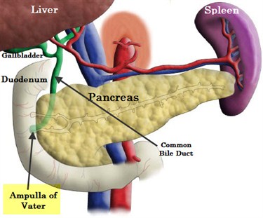
Ampullary cancer is a malignant tumor that arises from the Ampulla of Vater, the last centimeter of the common bile duct as it passes through the duodenum, the first section of the intestine. All pancreatic and biliary secretions enter the duodenum through the Ampulla of Vater.
Ampulla of Vater
A tumor blocking  the Ampulla of Vater will interfere with drainage of the pancreatic and biliary secretions into the intestine. Jaundice results when the drainage of bile into the duodenum is blocked causing it to accumulate in in the bloodstream. Jaundice, the yellowing of the skin, is typically one of the first symptoms present with Ampullary cancer.
The diagnostic tests used to for ampullary cancer  are similar to those for pancreatic cancer; endoscopy or endoscopic retrograde cholangiopancreatography (ERCP) are frequently used to make the diagnosis. 


                        Histology of Periampullay adenocarcinoma


Patients with periampullary cancer who received chemotherapy and surgery lived longer than patients who did not receive the chemotherapy.
This is the finding of a large multi-center research effort that was published in the July 11 issue of JAMA (Journal of the American Medical Association).
This an important large international multi-center study showing the benefit of chemotherapy after surgical resection of a specific type ofperiampullary cancer," James Farrell, MD, director of the University of California Los Angeles Medical Center Endoscopic Ultrasound Division of Digestive Diseases, told dailyRx in an email,
"It supports the use of adjuvant treatment with gemcitabine for this group of patients," Dr. Farrell said.
Additional study is needed to learn more about this treatment option. "There were different survival outcomes by tumor type, although age, poorly differentiated tumor grade, and lymph node involvement were also independent survival factors,” the authors write.

Tuesday, June 26, 2012

A Rare Cancer in Children

It is always heartbreaking when children have cancer. Usually cancer in children affects the blood cells or brain. A very rare cancer in children is rhabdomyosarcoma. Where is this cancer located in a child's body?

Fewer than 60 children are diagnosed with rhabdomyosarcoma in the UK each year. About the same number in the United States. Most of them are younger than 10 years old. It's more common in boys than girls.
Rhabdomyosarcoma is the most common of the soft tissue sarcomas in children. These tumors develop from muscle or fibrous tissue and can grow in any part of the body.
The most common areas of the body to be affected are around the head and neck, the bladder or the testes. Sometimes tumours are also found in a muscle or a limb, in the chest or in the abdominal wall. If the tumour is in the head or neck region, it can occasionally spread into the brain or the fluid around the spinal cord.

What causes this disease is unknown. Children who have rare genetic disorders are more prone to have rhabdommyosarcoma.

                               Image of rhabdomyosarcoma that has been removed from a child's body.

The images of the children with this cancer are very disturbing so they will not be displayed.

The signs and symptoms will depend on the part of the body that's affected by the rhabdomyosarcoma. The most common sign is a swelling or lump.
  • If the tumor is in the head area, it can sometimes cause a blockage (obstruction) and a discharge from the nose or throat. Occasionally, an eye may appear swollen and protruding.
  • If the tumor is in the abdomen (tummy), your child may have discomfort in the abdomen and difficulty going to the toilet.
  • If the tumor is in the bladder, your child may have blood in the urine and difficulty passing urine.  
A variety of tests and investigations may be needed to diagnose a rhabdomyosarcoma. A small operation may be needed to remove a sample from the tumour to be looked at under a microscope. This called a biopsy. It's usually done under a general anaesthetic.
Various tests may be done to check the exact size of the tumour and whether it has spread to any other part of the body. These may include:
  • a chest x-ray to check the lungs
  • an ultrasound
  • CT or MRI scans
  • blood and bone marrow tests.
Any tests and investigations that your child needs will be explained to you. The booklet A parent's guide to children's cancer gives details of what the tests and scans involve.

Rhabdomyosarcomas are rare tumours and should be treated at specialist centres. 
Treatment depends upon the size of the tumour, its position within the body, and whether it has spread. Treatment of rhabdomyosarcoma usually includes surgery, radiotherapy or chemotherapy, or a combination of these treatments.

Surgery

If at all possible, surgery will be used to remove the tumour. Chemotherapy, using a combination of drugs, is often given before surgery to shrink the tumour. Radiotherapy may also be given to the area of the tumour, particularly if it cannot be completely removed by surgery.

Chemotherapy

If the tumour cannot be removed with surgery, treatment will usually involve a combination of chemotherapy and radiotherapy. Chemotherapy is the use of anti-cancer (cytotoxic) drugs to destroy cancer cells and is usually given every three weeks. It may be given to shrink the tumour before surgery or with radiotherapy when the tumour can't be removed by surgery. The drugs used and the length of treatment depends on the type and stage of the rhabdomyosarcoma.

Radiotherapy

Radiotherapy treats cancer by using high-energy rays, which destroy the cancer cells while doing as little harm as possible to normal cells. It's given to the area where the rhabdomyosarcoma occurs.

Side effects of treatment 

Treatment for rhabdomyosarcoma often causes side effects, and your child’s doctor will discuss this with you before treatment starts. Any possible side effects will depend on the particular treatment being given and the part of the body that's being treated.
Chemotherapy can make your child feel better by relieving the symptoms of the cancer, but it can sometimes have side effects such as feeling sick (nausea) and being sick (vomiting), hair loss, an increased risk of infection, bruising and bleeding, tiredness and diarrhoea.

Late side effects

A small number of children may develop side effects many years after their treatment for a rhabdomyosarcoma. Long-term side effects depend on the type of treatment used, and may include a possible reduction in bone growth, infertility, a change in the way the heart and the kidneys work, and a slight increase in the risk of developing another cancer in later life.
Your child’s doctor or nurse will talk to you about any possible late side effects. There is more detailed information about these late side effects in the booklet A parent’s guide to children’s cancer.

                                        Mayo Clinic Oncologist Explains Rhabdomyosarcoma


                                       Holly's Journey Dealing With This Rare Cancer

http://www.macmillan.org.uk/Cancerinformation/Cancertypes/Childrenscancers/Typesofchildrenscancers/Rhabdomyosarcoma.aspx

Monday, June 25, 2012

New Biomarkers Tests for Ovarian Cancer

Great new article about new biomarker testing at the end of this year concerning ovarian cancer. Every woman needs to read this to educate yourself and share with others about what is involved with this life threatening cancer.

Autotelic Lab, Fountain Valley, a developer of quantitative rapid tests, announced today that it will be presenting a poster “The Hormones BNP and FSH in Ovarian Cancer: Potential as Diagnostic Biomarkers” at ENDO 2012. Ovarian cancer accounts for approximately 3 percent of all cancers in women and is the fifth leading cause of cancer-related death among women in the United States. Ovarian cancer has the highest mortality of all cancers of the female reproductive system. This reflects, in part, a lack of early symptoms and effective ovarian cancer screening tests. Thus, ovarian cancer is often diagnosed at an advanced stage, after the cancer has spread beyond the ovary. Cancer Antigen 125 (CA125, aka Mucin16) is overproduced by ovarian cancer cells; however, it cannot be used as a diagnostic biomarker for ovarian cancer as it can be absent when disease is present, or levels can be high when no disease or no malignant disease exists. Serial changes in CA125 levels, if elevated, however, can be useful in assessment of disease status and progression. Ovarian epithelial cancer is more common in individuals with elevated Gonadotropin-releasing hormones (GnRH) including Follicle Stimulating Hormone (FSH) and Luteinizing Hormone (LH) such as postmenopausal women or women who have received treatment to induce ovulation. Conversely, reduced risk of ovarian cancer is associated with a history of multiple pregnancies, breastfeeding, oral contraceptive use, and estrogen replacement therapy, all of which are related to lower levels of and reduced exposure to FSH and LH. FSH regulates gene expression in ovarian tumors and causes neovascularization of ovarian cancers by increasing Vascular Endothelial Growth Factor (VEGF) expression through upregulation of survivin. We therefore examined FSH levels in relation to CA125 as potential diagnostic marker for ovarian cancer using a quantitative point-of-care device for FSH that was recently developed for therapeutic drug monitoring. The surprising potential of FSH and/or Brain Natriuretic Peptide (BNP) as diagnostic biomarkers for ovarian cancer will be presented.

These tests are performed on serum from a blood draw. It usually take a few days to get the results,but are very informative for the oncologist as far as plan of treatment.


                                  An Inspiring Story of a woman (physician) Diagnosed With Ovarian

Cancerhttp://news.topwirenews.com/2012/06/23/fsh-and-bnp-as-potential-diagnostic-biomarker-for-ovarian-cancer/%

Monday, June 18, 2012

Blood Test and Early Stage Breast Cancer

 Through cancer research it has been determined that a current blood test can determine which patients who were diagnosed with early stages of breast cancer and who had their tumors surgically removed may see their cancer return.
The following study will explain how this was determined.

Doctors at the M.D. Anderson Cancer Center in Houston, Texas, took blood samples from 302 women with stage 1, 2 or 3 breast cancer right before they had their tumor surgically removed.  None of the women had been treated with chemotherapy. The patients' progress was followed for nearly 3 years (35 months).
According to the study published Tuesday in the journal Lancet Oncology , 25% of women whose cancer was confined to the breast had at least one circulating tumor cell in their 7.5 milliliter sample of blood (about the equivalent of half a tablespoon).  Normally a lymph node biopsy is used to determine the likelihood of the cancer coming back.
Researchers found 73 women (24%) had at least one circulating tumor cell; 29 women (about 10%) had at least two circulating tumor cells in their sample and 16 (5%) had at least 3 or more tumor cells in their blood sample.
Women with early stage breast cancer aren't usually thought to be at high risk for having their cancer come back, but some do and doctors don't know why, which is why lead study author Dr. Anthony Lucci says he and his colleagues decided to investigate this.
The study found that if a women had just one circulating tumor cell in her sample, she had a 4 times greater chance of dying, compared to a woman with no CTCs.
"If three or more tumor cells were found in a blood sample, the woman had an almost 11 times higher risk of dying from the cancer compared to those who didn't have any circulating tumor cells," says Lucci.
All in all, 31% of the women in this study found their cancer came back or died during the study period.
Lucci likens circulating tumor cells to seeds in a garden. If you dump a bag of seeds in the dirt, he says, the odds that one or several take hold and grow is much higher than planting just one seed.


                                                           Circulating Tumor Cells
"While the findings of an association between circulating tumor cells  and outcome in breast cancer is not novel," says Dr. Boris Pasche, Director of the Division of Hemotology and Oncology at the University of Alabama at Birmingham.  "The strength of this new paper is its capability to characterize the magnitude of the impact of CTCs on breast cancer outcome."   He says the other strength of this study is that the blood samples were taken just before surgery, giving a more accurate picture of where traces of cancer are outside the breast.
Once a tumor is removed, it's possible for cells to break off, thus skewing the accuracy of how much cancer is already circulating outside the breast.
According to a commentary in the Journal of the American Medical Association in March 2010 , the first documented research on circulating tumors was conducted in 1846. Since then, studies have shown that looking for circulating tumor cells in patients, who've already seen their cancer spread, can be an important tool to determine how a patient does in the future.
This hasn't been proven to be the case for early stage breast cancer, where the cancer has not spread beyond the breast and it is something this new study doesn't do either.
However, Pasche, who was not involved with the research, says if the results are validated in larger clinical trials, this may lead to a new screening method for predicting which women may be more likely to see their cancer come back.
"This provides a new train of thought of how we should handle women with early-stage breast cancer because we could identify the women who have a high risk of recurrences and early death and treat them differently."   Pasche suggests those patients  get more aggressive removal of lymph nodes, they may be put on chemotherapy (something they may not normally be prescribed) and their doctors will probably have a much tighter follow-up schedule for them.
Lucci and Pasche say this type of screening is not yet ready for prime time, and M.D. Anderson already has further studies underway.
                                                         Circulating Tumor Cells and  Breast Cancer Recurrencehttp://www.wtnh.com/dpps/health/cancer/blood-test-may-signal-breast-cancers-return_4198976#.T9-D67UTpJs