Showing posts with label images. Show all posts

Tuesday, March 20, 2012

Melanoma Of The Eye

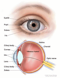
When melanoma is discussed we immediately think of cancer of the skin , but it also can be present in our eyes. This type of cancer is called Intraocular Melanoma.


                                                               Melanoma in the iris of the eye.

                                                            Melanoma in the retina of the eye


Intraocular melanoma begins in the middle of 3 layers of the wall of the eye. The outer layer includes the white sclera (the "white of the eye") and the clear cornea at the front of the eye. The inner layer has a lining of nerve tissue, called the retina, which senses light and sends images along the optic nerve to the brain.

This type of cancer most often occurs in people who are middle aged. In most cases of intraocular melanoma, doctors detect the cancer during a routine eye examination. The chance of recovery (prognosis) will depend on factors such as the size and cell type of the cancer. This type of melanoma is rare.

Most people with intraocular melanoma experience no symptoms of the cancer in its early stages. Melanoma that starts in the iris may appear as a dark spot on the iris. Intraocular melanoma that is in the ciliary body or choroid may cause blurry vision.

Age and sun exposure may increase the risk of developing intraocular melanoma.
Anything that increases your risk of getting a disease is called a risk factor. Having a risk factor does not mean that you will get cancer; not having risk factors doesn’t mean that you will not get cancer. People who think they may be at risk should discuss this with their doctor. Risk factors for intraocular melanoma include the following:
  • Older age
  • Being white
  • Having a fair complexion (light skin) or green or blue eyes.
  • Being able to tan
Possible signs of intraocular melanoma include a dark spot on the iris or blurred vision.
Intraocular melanoma may not cause any early symptoms. It is sometimes found during a routine eye exam when the doctor dilates the pupil and looks into the eye. The following symptoms may be caused by intraocular melanoma or by other conditions. A doctor should be consulted if any of these problems occur:
  • A dark spot on the iris
  • Blurred vision
  • A change in the shape of the pupil
  • A change in vision
Glaucoma may develop if the tumor causes the retina to separate from the eye. If this happens, there may be no symptoms, or symptoms may include the following:
  • Eye pain
  • Blurred vision
  • Eye redness
  • Nausea
 

Doctors stage intraocular melanoma based on the area of the eye where the tumor is found and the size of the tumor. The stages of intraocular melanoma include:
 
  • Iris melanoma
  • Ciliary body melanoma
  • Small choroidal melanoma
  • Medium and large choroidal melanoma
  • Extraocular extension and metastatic intraocular melanoma
  • Recurrent intraocular melanoma. 
  •  


Iris Melanoma
Intraocular melanoma of the iris occurs in the front colored part of the eye. Iris melanomas usually grow slowly and do not spread to other parts of the body.
 
Ciliary Body Melanoma
Intraocular melanoma of the ciliary body occurs in the back part of the eye.
 
Small Choroidal Melanoma
Intraocular melanoma of the choroid occurs in the back part of the eye. This type of tumor is classified by the size of the tumor. A small choroidal melanoma is 3 millimeters or less in thickness.
 
Medium and Large Choroidal Melanoma
Intraocular melanomas of the choroid occur in the back part of the eye. This type of tumor is classified by the size of the tumor. Medium and large choroidal melanomas are more than 3 millimeters in thickness.
 
Extraocular Extension and Metastatic Intraocular Melanoma
In extraocular extension and metastatic intraocular melanoma, the melanoma has spread outside the eye, to the nerve behind the eye (the optic nerve), to the eye socket, or to other parts of the body.
 
Recurrent
Recurrent intraocular melanoma refers to cases of the cancer that have come back (recurred) after they were treated.
 

Treatment for Intraocular Melanoma

Treatment options for intraocular melanoma may include:
 
  • Surgery (taking out the cancer)
  • Radiation therapy (using high-dose x-rays or other high-energy rays to kill cancer cells)
  • Laser therapy (using an intensely powerful beam of light to destroy the tumor or blood vessels that feed the tumor).
 
In some cases (such as when the cancer is small and causing no symptoms), the treatment plan may involve monitoring the patient's cancer carefully and waiting to treat it until it changes or causes symptoms. This is sometimes known as watchful waiting.  

                                           Video of a cancerous tumor of eye surgically removed.

http://my.clevelandclinic.org/disorders/intraocular_melanoma/hic_intraocular_melanoma.aspx

http://skin-cancer.emedtv.com/intraocular-melanoma/intraocular-melanoma-p3.html

Thursday, March 1, 2012

What Is Bladder Cancer And How Is It Diagnosed?

Bladder cancer begins in the cells that line the inside of the bladder.  The bladder is the place in your lower abdomen that stores the urine.  Bladder cancer usually affects older patients , but anyone of any age can have this type of cancer.


                                                Bladder tumor

The great majority of bladder cancers are diagnosed at an early stage — when bladder cancer is highly treatable. However, even early-stage bladder cancer is likely to recur. For this reason, bladder cancer survivors often undergo follow-up tests to look for bladder cancer recurrence for years after treatment.

Diagnosing bladder cancer
Tests and procedures used to diagnose bladder cancer may include:
  • Cystoscopy. During cystoscopy, your doctor inserts a narrow tube (cystoscope) through your urethra. The cystoscope has a lens and fiber-optic lighting system, allowing your doctor to see the inside of your urethra and bladder. You usually receive a local anesthetic during cystoscopy to make you more comfortable.
  • Biopsy. During cystoscopy, your doctor may pass a special tool through your urethra and into your bladder in order to collect a small cell sample (biopsy) for testing. This procedure is sometimes called transurethral resection of bladder tumor (TURBT). TURBT can also be used to treat bladder cancer. TURBT is usually performed under general anesthesia.
  • Urine cytology. A sample of your urine is analyzed under a microscope to check for cancer cells in a procedure called urine cytology.
  • Imaging tests. Imaging tests allow your doctor to examine the structures of your urinary tract. You may receive a dye, which can be injected into a vein. An intravenous pyelogram is a type of X-ray imaging test that uses a dye to highlight your kidneys, ureters and bladder. A computerized tomography (CT) scan is a type of X-ray test that allows your doctor to better see your urinary tract and the surrounding tissues.
Staging bladder cancer
Once it's confirmed that you have bladder cancer, your doctor may order additional tests to determine the extent, or stage, of the cancer. Staging tests may include:
  • CT scan
  • Magnetic resonance imaging (MRI)
  • Bone scan
  • Chest X-ray
Bladder cancer stages
The stages of bladder cancer are:
  • Stage I. Cancer at this stage occurs in the bladder's inner lining, but hasn't invaded the muscular bladder wall.
  • Stage II. At this stage, cancer has invaded the bladder wall, but is still confined to the bladder.
  • Stage III. The cancer cells have spread through the bladder wall to surrounding tissue. They may also have spread to the prostate in men or the uterus or vagina in women.
  • Stage IV. By this stage, cancer cells may have spread to the lymph nodes and other organs, such as your lungs, bones or liver.
                                               Mayo Clinic Discussion of Bladder Cancer

  • For more information: http://www.ncbi.nlm.nih.gov/pubmedhealth/PMH0001517/ or
  • http://www.mayoclinic.com/health/bladder-cancer/DS00177/DSECTION=tests%2Dand%2Ddiagnosis


Saturday, February 25, 2012

The Facts About Bone Cancer

Did you know that bone cancer rarely is the only location that cancer is seen in the human body? Usually it is spread or metastasized by remote cancers in other organs of the body.

                                              Bone tumor by X-Ray


                                            Bone Cancer under the microscope from biopsy

Notice the abnormal growth of cells in the picture above which indicates cancer.

The Most Common Types of Bone Cancers :

  • Osteosarcoma, which arises from osteoid tissue in the bone. This tumor occurs most often in the knee and upper arm (1).
  • Chondrosarcoma, which begins in cartilaginous tissue. Cartilage pads the ends of bones and lines the joints. Chondrosarcoma occurs most often in the pelvis (located between the hip bones), upper leg, and shoulder. Sometimes a chondrosarcoma contains cancerous bone cells. In that case, doctors classify the tumor as an osteosarcoma.
  • The Ewing Sarcoma Family of Tumors (ESFTs), which usually occur in bone but may also arise in soft tissue (muscle, fat, fibrous tissue, blood vessels, or other supporting tissue). Scientists think that ESFTs arise from elements of primitive nerve tissue in the bone or soft tissue (2). ESFTs occur most commonly along the backbone and pelvis and in the legs and arms (3).
Other types of cancer that arise in soft tissue are called soft tissue sarcomas. They are not bone cancer and are not described in this resource.

What are bone cancer symptoms and signs?

The most common symptom of bone tumors is pain. In most cases, the symptoms become gradually more severe with time, including bone pain. Initially, the pain may only be present either at night or with activity. Depending on the growth of the tumor, those affected may have symptoms for weeks, months, or years before seeking medical advice. In some cases, a mass or lump may be felt either on the bone or in the tissues surrounding the bone. This is most common with MFH or fibrosarcoma but can occur with other bone tumors. The bones can become weakened by the tumor and lead to a fracture after little or no trauma or just from standing on the affected bone. This can occur with both benign and malignant tumors. Even benign tumors can spread locally and weaken the surrounding bone. If the tumor compresses the surrounding nerve it can cause pain, numbness, or tingling in the extremities. If the surrounding blood vessels are compressed, it can affect the blood flow to the extremities. Fever, chills, night sweats, and weight loss can occur but are less common. These symptoms are more common after spread of the tumor to other tissues in the body.

What are the possible causes of bone cancer?

Although bone cancer does not have a clearly defined cause, researchers have identified several factors that increase the likelihood of developing these tumors. Osteosarcoma occurs more frequently in people who have had high-dose external radiation therapy or treatment with certain anticancer drugs; children seem to be particularly susceptible. A small number of bone cancers are due to heredity. For example, children who have had hereditary retinoblastoma (an uncommon cancer of the eye) are at a higher risk of developing osteosarcoma, particularly if they are treated with radiation. Additionally, people who have hereditary defects of bones and people with metal implants, which doctors sometimes use to repair fractures, are more likely to develop osteosarcoma (4). Ewing sarcoma is not strongly associated with any heredity cancer syndromes, congenital childhood diseases, or previous radiation exposure (2).

What are the treatment options for bone cancer?

Treatment options depend on the type, size, location, and stage of the cancer, as well as the person’s age and general health. Treatment options for bone cancer include surgery, chemotherapy, radiation therapy, and cryosurgery.
  • Surgery is the usual treatment for bone cancer. The surgeon removes the entire tumor with negative margins (no cancer cells are found at the edge or border of the tissue removed during surgery). The surgeon may also use special surgical techniques to minimize the amount of healthy tissue removed with the tumor.
    Dramatic improvements in surgical techniques and preoperative tumor treatment have made it possible for most patients with bone cancer in an arm or leg to avoid radical surgical procedures (removal of the entire limb). However, most patients who undergo limb-sparing surgery need reconstructive surgery to maximize limb function (1).
  • Chemotherapy is the use of anticancer drugs to kill cancer cells. Patients who have bone cancer usually receive a combination of anticancer drugs. However, chemotherapy is not currently used to treat chondrosarcoma (1).
  • Radiation therapy, also called radiotherapy, involves the use of high-energy x-rays to kill cancer cells. This treatment may be used in combination with surgery. It is often used to treat chondrosarcoma, which cannot be treated with chemotherapy, as well as ESFTs (1). It may also be used for patients who refuse surgery.
  • Cryosurgery is the use of liquid nitrogen to freeze and kill cancer cells. This technique can sometimes be used instead of conventional surgery to destroy the tumor (1).


    This video educates us on how bone cancer is diagnosed.  To learn more about bone cancer then go to the following websites :

    http://www.cancer.gov/cancertopics/factsheet/Sites-Types/bone

    http://www.medicinenet.com/bone_cancer/page3.htm