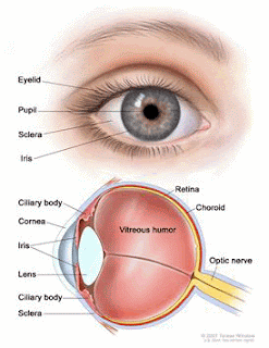
Melanoma in the iris of the eye.
Melanoma in the retina of the eye
Intraocular melanoma begins in the middle of 3 layers of the wall of the eye. The outer layer includes the white sclera (the "white of the eye") and the clear cornea at the front of the eye. The inner layer has a lining of nerve tissue, called the retina, which senses light and sends images along the optic nerve to the brain.
This type of cancer most often occurs in people who are middle aged. In most cases of intraocular melanoma, doctors detect the cancer during a routine eye examination. The chance of recovery (prognosis) will depend on factors such as the size and cell type of the cancer. This type of melanoma is rare.
Most people with intraocular melanoma experience no symptoms of the cancer in its early stages. Melanoma that starts in the iris may appear as a dark spot on the iris. Intraocular melanoma that is in the ciliary body or choroid may cause blurry vision.
Age and sun exposure may increase the risk of developing intraocular melanoma.
Anything that increases your risk of getting a disease is called a risk factor. Having a risk factor does not mean that you will get cancer; not having risk factors doesn’t mean that you will not get cancer. People who think they may be at risk should discuss this with their doctor. Risk factors for intraocular melanoma include the following:
- Older age
- Being white
- Having a fair complexion (light skin) or green or blue eyes.
- Being able to tan
Intraocular melanoma may not cause any early symptoms. It is sometimes found during a routine eye exam when the doctor dilates the pupil and looks into the eye. The following symptoms may be caused by intraocular melanoma or by other conditions. A doctor should be consulted if any of these problems occur:
- A dark spot on the iris
- Blurred vision
- A change in the shape of the pupil
- A change in vision
- Eye pain
- Blurred vision
- Eye redness
- Nausea
Doctors stage intraocular melanoma based on the area of the eye
where the tumor is found and the size of the tumor. The stages of
intraocular melanoma include:
- Iris melanoma
- Ciliary body melanoma
- Small choroidal melanoma
- Medium and large choroidal melanoma
- Extraocular extension and metastatic intraocular melanoma
- Recurrent intraocular melanoma.
Iris Melanoma
Intraocular melanoma of the iris occurs in the front colored part
of the eye. Iris melanomas usually grow slowly and do not spread to
other parts of the body.
Ciliary Body Melanoma
Intraocular melanoma of the ciliary body occurs in the back part of the eye.
Small Choroidal Melanoma
Intraocular melanoma of the choroid occurs in the back part of the
eye. This type of tumor is classified by the size of the tumor. A small
choroidal melanoma is 3 millimeters or less in thickness.
Medium and Large Choroidal Melanoma
Intraocular melanomas of the choroid occur in the back part of the
eye. This type of tumor is classified by the size of the tumor. Medium
and large choroidal melanomas are more than 3 millimeters in thickness.
Extraocular Extension and Metastatic Intraocular Melanoma
In extraocular extension and metastatic intraocular melanoma, the
melanoma has spread outside the eye, to the nerve behind the eye (the
optic nerve), to the eye socket, or to other parts of the body.
Recurrent
Recurrent intraocular melanoma refers to cases of the cancer that have come back (recurred) after they were treated.
Treatment options for intraocular melanoma may include:
- Surgery (taking out the cancer)
- Radiation therapy (using high-dose x-rays or other high-energy rays to kill cancer cells)
- Laser therapy (using an intensely powerful beam of light to destroy the tumor or blood vessels that feed the tumor).
Video of a cancerous tumor of eye surgically removed.
http://my.clevelandclinic.org/disorders/intraocular_melanoma/hic_intraocular_melanoma.aspx
http://skin-cancer.emedtv.com/intraocular-melanoma/intraocular-melanoma-p3.html


No comments:
Post a Comment